. 2001 Apr;21(7):2521-32.
doi: 10.1128/MCB.21.7.2521-2532.2001.

Essential role of insulin receptor substrate 1 (IRS-1) and IRS-2 in adipocyte differentiation

Affiliations

Essential role of insulin receptor substrate 1 (IRS-1) and IRS-2 in adipocyte differentiation

H Miki et al. Mol Cell Biol. 2001 Apr.

Abstract

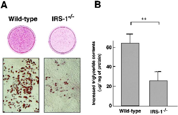
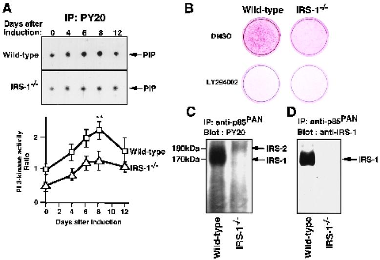
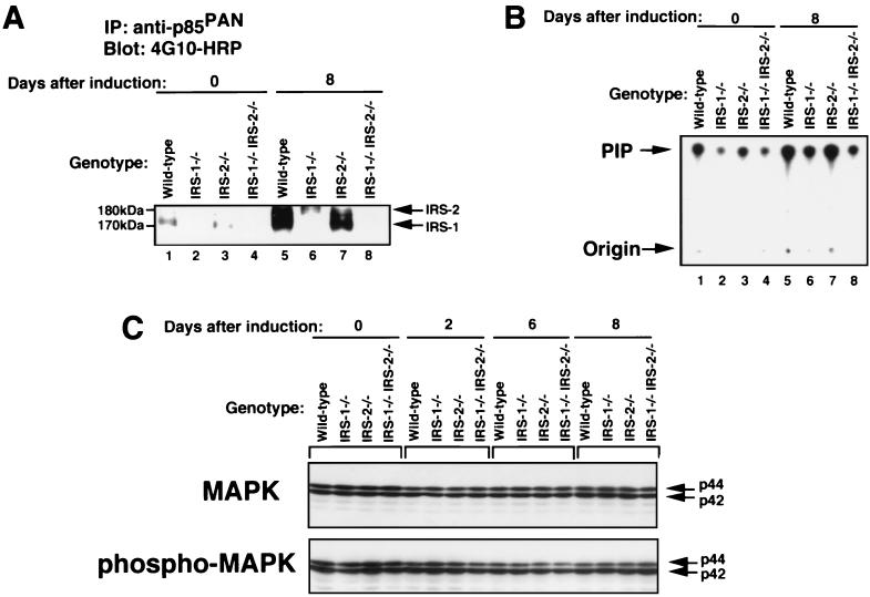
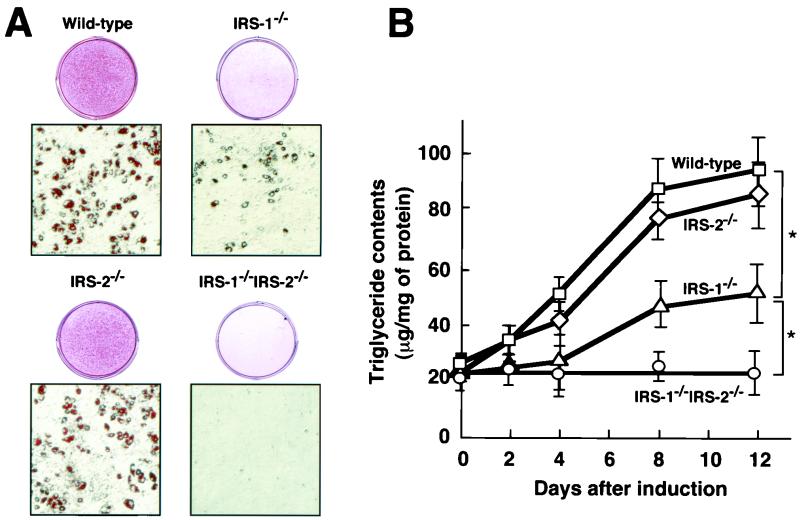
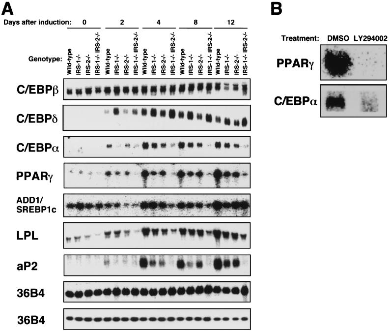
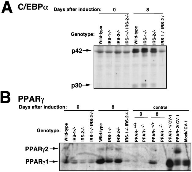
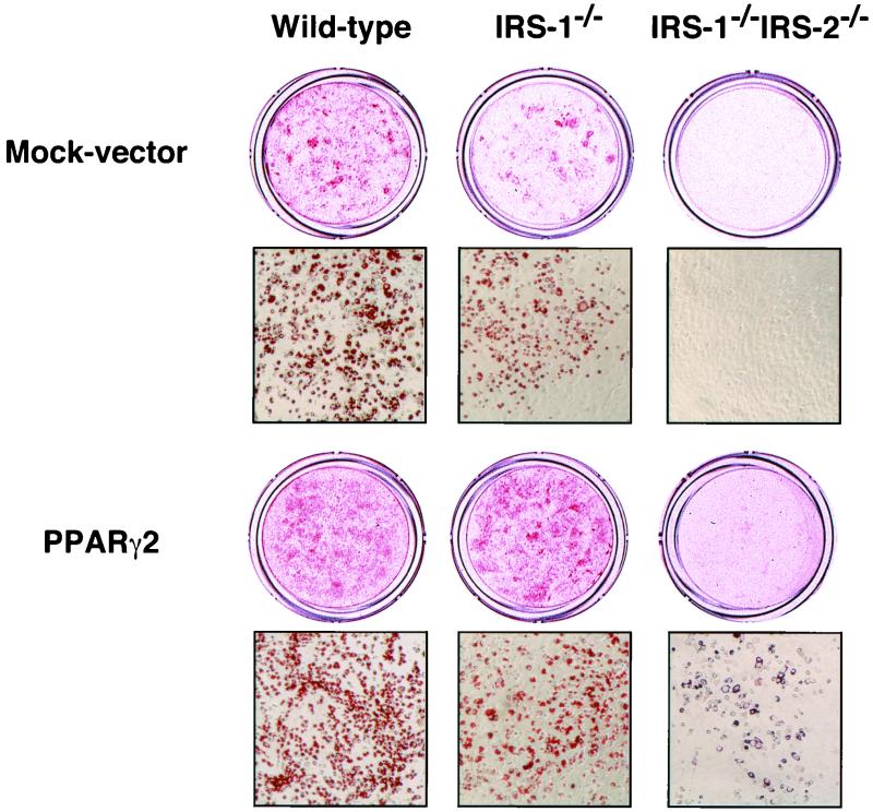
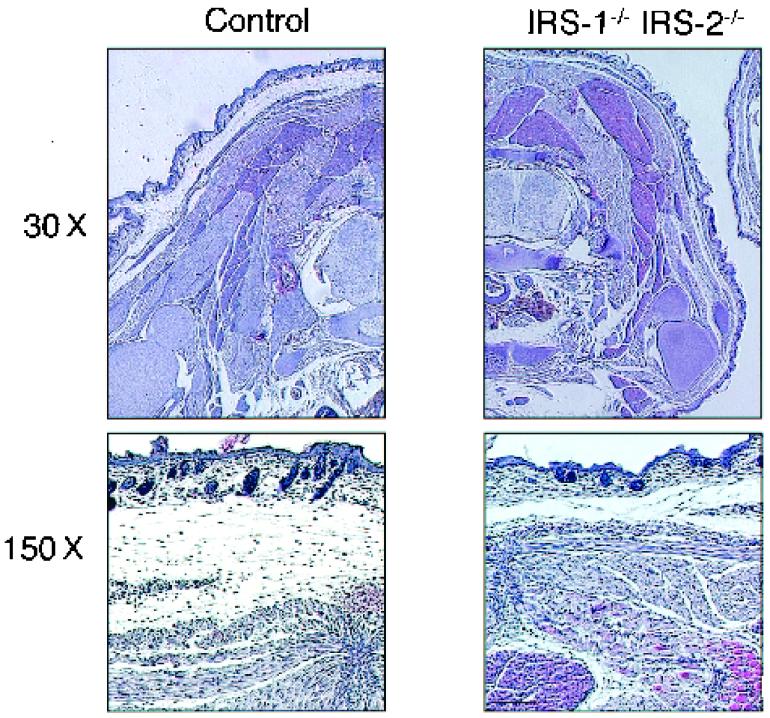
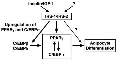
To investigate the role of insulin receptor substrate 1 (IRS-1) and IRS-2, the two ubiquitously expressed IRS proteins, in adipocyte differentiation, we established embryonic fibroblast cells with four different genotypes, i.e., wild-type, IRS-1 deficient (IRS-1(-/-)), IRS-2 deficient (IRS-2(-/-)), and IRS-1 IRS-2 double deficient (IRS-1(-/-) IRS-2(-/-)), from mouse embryos of the corresponding genotypes. The abilities of IRS-1(-/-) cells and IRS-2(-/-) cells to differentiate into adipocytes are approximately 60 and 15%, respectively, lower than that of wild-type cells, at day 8 after induction and, surprisingly, IRS-1(-/-) IRS-2(-/-) cells have no ability to differentiate into adipocytes. The expression of CCAAT/enhancer binding protein alpha (C/EBPalpha) and peroxisome proliferator-activated receptor gamma (PPARgamma) is severely decreased in IRS-1(-/-) IRS-2(-/-) cells at both the mRNA and the protein level, and the mRNAs of lipoprotein lipase and adipocyte fatty acid binding protein are severely decreased in IRS-1(-/-) IRS-2(-/-) cells. Phosphatidylinositol 3-kinase (PI 3-kinase) activity that increases during adipocyte differentiation is almost completely abolished in IRS-1(-/-) IRS-2(-/-) cells. Treatment of wild-type cells with a PI 3-kinase inhibitor, LY294002, markedly decreases the expression of C/EBPalpha and PPARgamma, a result which is associated with a complete block of adipocyte differentiation. Moreover, histologic analysis of IRS-1(-/-) IRS-2(-/-) double-knockout mice 8 h after birth reveals severe reduction in white adipose tissue mass. Our results suggest that IRS-1 and IRS-2 play a crucial role in the upregulation of the C/EBPalpha and PPARgamma expression and adipocyte differentiation.


Figures

FIG. 1
FIG. 1
Ability of wild-type and IRS-1−/− cells to differentiate into adipocytes. (A) Oil-Red O staining for fat accumulation in wild-type and IRS-1−/− cells at day 8 after induction. Cells were grown to confluence and then induced to differentiation by exposure to IBMX, DEX, and insulin, as described in Materials and Methods. (B) Increase in intracellular triglyceride content from day 0 to day 8 after induction. The assays were performed as described in Materials and Methods. The data represent the means ± the standard errors of the means from four experiments. Wild-type, n = 6; IRS-1−/−, n = 6; ∗∗, P < 0.01.
FIG. 2
FIG. 2
Role of PI 3-kinase in adipocyte differentiation of wild-type and IRS-1−/− cells. (A) PI 3-kinase activities during adipocyte differentiation. The antiphosphotyrosine antibody (PY20) immunoprecipitates from 50 μg of protein of cellular lysates of wild-type and IRS-1−/− cells at the day indicated after induction into adipocytes were subjected to a PI 3-kinase assay. The autoradiograms of the spots corresponding to PIP are shown in the upper panel. The radioactivity in the spots was measured, and the results are shown in the lower panel, expressed as the ratios of the values for the respective cells to those for wild-type cells at day 0 after induction. The data represent the means ± the standard errors of the means from three independent experiments. ∗∗, P < 0.01. (B) Effect of the PI 3-kinase inhibitor LY294002 on adipocyte differentiation of wild-type and IRS-1−/− cells. LY294002 (20 μM) was added to adipocyte differentiation medium, and the medium was changed at 36-h intervals. As a control, 0.1% dimethyl sulfoxide was added to the medium, and the medium was changed at the same intervals. At day 8 after induction, the intracellular fat accumulation was assessed by staining with Oil-Red O. (C) Tyrosine-phosphorylated proteins associated with p85 subunit of PI 3-kinase. Total lysates (100 μg of protein each) of cells at day 8 after induction were immunoprecipitated with anti-p85PAN (antibody to the p85 subunit of PI 3-kinase), and the immunoprecipitates were subjected to SDS-PAGE followed by immunoblotting with PY20 conjugated to HRP. (D) Association of IRS-1 with p85 subunit of PI 3-kinase. Total lysates (100 μg of protein each) of cells at day 8 after induction were immunoprecipitated with anti-p85PAN, and the immunoprecipitates were subjected to SDS-PAGE, followed by immunoblotting with anti-IRS-1 antibody.
FIG. 3
FIG. 3
PI 3-kinase and MAPK activities during adipocyte differentiation in wild-type, IRS-1−/−, IRS-2−/−, and IRS-1−/− IRS-2−/− cells. (A) Tyrosine-phosphorylated proteins associated with p85 subunit of PI 3-kinase in cells with four different genotypes, wild-type, IRS-1−/−, IRS-2−/−, and IRS-1−/− IRS-2−/− cells. Total lysates (100 μg of protein each) of cells at day 0 and day 8 after induction were immunoprecipitated with anti-p85PAN (antibody to the p85 subunit of PI 3-kinase), and the immunoprecipitates were subjected to SDS-PAGE, followed by immunoblotting with antiphosphotyrosine antibody (4G10) conjugated to HRP. (B) PI 3-kinase activity associated with tyrosine phosphorylated proteins in wild-type, IRS-1−/−, IRS-2−/−, and IRS-1−/− IRS-2−/− cells. The immunoprecipitates with PY20 from cell lysates (50 μg of protein) were subjected to the PI 3-kinase assay. The autoradiogram of the thin-layer chromatograph is shown. (C) Levels of MAPK protein (upper panel) and phosphorylation of MAPK (lower panel) during adipocyte differentiation. Total lysates (10 μg of protein each) of cells with the four different genotypes were subjected to immunoblotting as described in Materials and Methods using antibody specific to either MAPK or phospho-MAPK.
FIG. 4
FIG. 4
Adipocyte differentiation of EF cells with the four different genotypes, i.e., wild-type, IRS-1−/−, IRS-2−/−, and IRS-1−/− IRS-2−/− cells. (A) Oil-Red O staining for fat accumulation in cells with the four different genotypes. Wild-type, IRS-1−/−, IRS-2−/−, and IRS-1−/− IRS-2−/− cells were grown to confluence, exposed to IBMX, DEX, and insulin to induce differentiation as described in Materials and Methods and then stained with Oil-Red O at day 8 day after induction. (B) Intracellular triglyceride content during adipocyte differentiation of wild-type, IRS-1−/−, IRS-2−/−, and IRS-1−/− IRS-2−/− cells. The data represent the means ± the standard errors of the means from the analysis of wild type (n = 3), IRS-1−/− (n = 4), IRS-2−/− (n = 4), and IRS-1−/− IRS-2−/− (n = 3) cells. ∗, P < 0.05.
FIG. 5
FIG. 5
(A) Northern blotting analysis and RNase protection assay of transcriptional factors for adipocyte differentiation and adipogenic markers in wild-type, IRS-1−/−, IRS-2−/−, and IRS-1−/− IRS-2−/− cells during adipocyte differentiation. Total RNAs were extracted from adipocytes at days 0, 2, 4, 8, and 12 after induction, and a 6-μg sample of the total RNA was subjected to electrophoresis and hybridization with probes consisting of 32P-labeled cRNAs encoding C/EBPβ, C/EBPδ, C/EBPα, and 36B4 (upper panel) and with probes consisting of 32P-labeled cDNAs encoding aP2 and LPL. In an RNase protection assay, 10 μg of the total RNA was hybridized with cRNA encoding PPARγ, SREBP1c, and 36B4 (lower panel). 36B4 encodes acidic ribosomal phosphoprotein P0 and was used as a loading control. (B) Effect of the PI 3-kinase inhibitor LY294002 on expression of the mRNAs of PPARγ and C/EBPα in wild-type cells. LY294002 (20 μM) was added to adipocyte differentiation medium, and the medium was changed at 36-h intervals. As a control, 0.1% dimethyl sulfoxide was added to the medium, and the medium was changed at the same intervals. At day 8 after induction, total RNAs were extracted from wild-type cells, and a 6-μg sample of total RNA was electrophoresed and subjected to Northern blotting with PPARγ and C/EBPα.
FIG. 6
FIG. 6
Immunoblot analysis of C/EBPα (A) and PPARγ (B) in wild-type, IRS-1−/−, IRS-2−/−, and IRS-1−/− IRS-2−/− cells during adipocyte differentiation. Cells were grown to confluence and then exposed to IBMX, DEX, and insulin to induce differentiation. At days 0 and 8 after induction, the cells were lysed, and the lysates (6 μg of protein each) were subjected to SDS-PAGE, followed by immunoblotting with anti-C/EBPα antibody (A) and anti-PPARγ antibody (B) as described in Materials and Methods. In an immunoblot analysis of PPARγ(B), PPARγ-deficient cells described in a previous report (21) and the CV-1 cells in which PPARγ1 or PPARγ2 was overexpressed were also used as control samples.
FIG. 7
FIG. 7
Retrovirus-mediated reexpression of PPARγ improved the differentiation capacity of IRS-1−/− IRS-2−/− cells. Wild-type, IRS-1−/−, or IRS-1−/− IRS-2−/− cells were transfected with pMX-mPPARγ2-puro and mock vector by retrovirus-mediated gene transfer as described in Materials and Methods. At day 8 of differentiation, cells were stained with Oil-Red O to assess the intracellular fat accumulations.
FIG. 8
FIG. 8
Histological analyses of IRS-1−/− IRS-2−/− double-knockout mice showed severe reduction in WAT but not in BAT. Transverse sections at the level of the neck were made from ∼8-h control (left) and IRS-1−/− IRS-2−/− (right) mice. The sections were subjected to hematoxylin and eosin staining. Magnifications: ×30 (top) and ×150 (bottom).
FIG. 9
FIG. 9
Schematic model of the roles of IRS-1 and IRS-2 in adipogenesis. IRS-1 and IRS-2 play a crucial role in the upregulation of the expression of C/EBPα and PPARγ and adipocyte differentiation.

References

    1. Akira S, Isshiki H, Sugita T, Tanabe O, Kinoshita S, Nishio Y, Nakajima T, Hirano T, Kishimoto T. A nuclear factor for IL-6 expression (NF-IL6) is a member of a C/EBP family. EMBO J. 1990;9:1897–1906. - PMC - PubMed
    1. Altiok S, Xu M, Spiegelman B M. PPARγ induces cell cycle withdrawal: inhibition of E2F/DP DNA-binding activity via down-regulation of PP2A. Genes Dev. 1997;11:1987–1998. - PMC - PubMed
    1. Bruning J C, Winnay J, Cheatham B, Kahn C R. Differential signaling by insulin receptor substrate 1 (IRS-1) and IRS-2 in IRS-1-deficient cells. Mol Cell Biol. 1997;17:1513–1521. - PMC - PubMed
    1. Cao Z, Umek R M, McKnight S L. Regulated expression of three C/EBP isoforms during adipose conversion of 3T3–L1 cells. Genes Dev. 1991;5:1538–1552. - PubMed
    1. Catalioto R M, Gailard D, Ailhaud G, Negrel R. Terminal differentiation of mouse preadipocyte cells: the mitogenic-adipogenic role of growth hormone is mediated by the protein kinase C signalling pathway. Growth Factors. 1992;6:255–264. - PubMed

Publication types

Substances

LinkOut - more resources