. 2001 Mar 27;98(7):3685-9.
doi: 10.1073/pnas.071033998. Epub 2001 Mar 20.

The Sm domain is an ancient RNA-binding motif with oligo(U) specificity

Affiliations

The Sm domain is an ancient RNA-binding motif with oligo(U) specificity

T Achsel et al. Proc Natl Acad Sci U S A. .

Abstract

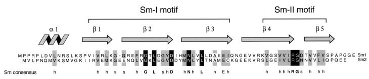
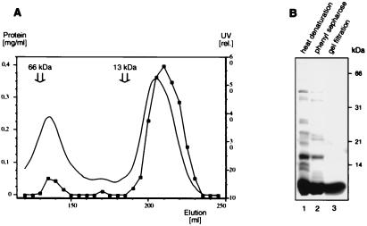
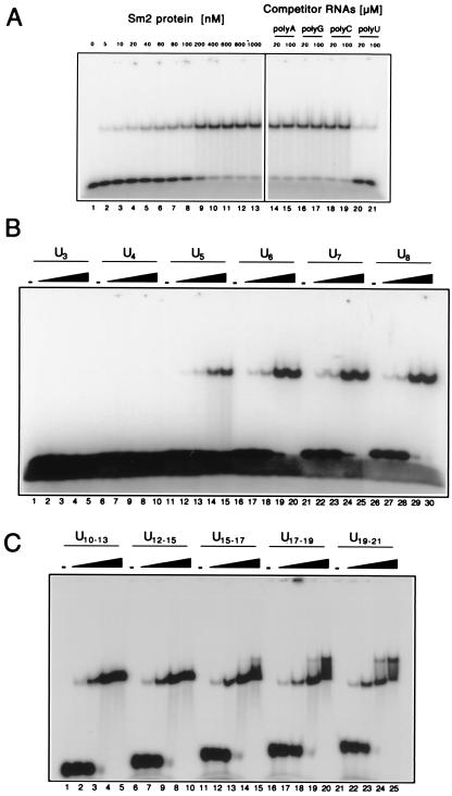
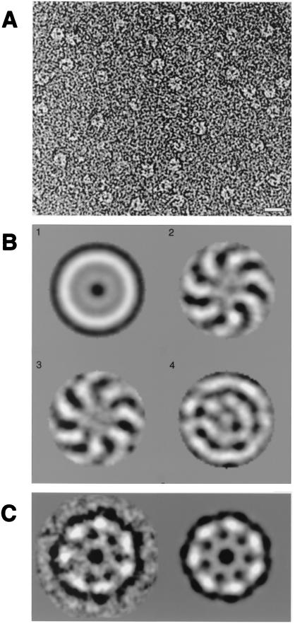
Sm and Sm-like proteins are members of a family of small proteins that is widespread throughout eukaryotic kingdoms. These proteins form heteromers with one another and bind, as heteromeric complexes, to various RNAs, recognizing primarily short U-rich stretches. Interestingly, completion of several genome projects revealed that archaea also contain genes that may encode Sm-like proteins. Herein, we studied the properties of one Sm-like protein derived from the archaebacterium Archaeoglobus fulgidus and overexpressed in Escherichia coli. This single small protein closely reflects the properties of an Sm or Sm-like protein heteromer. It binds to RNA with a high specificity for oligo(U), and assembles onto the RNA to form a complex that exhibits, as judged by electron microscopy, a ring-like structure similar to the ones observed with the Sm core ribonucleoprotein and the like Sm (LSm) protein heteromer. Importantly, multivariate statistical analysis of negative-stain electron-microscopic images revealed a sevenfold symmetry for the observed ring structure, indicating that the proteins form a homoheptamer. These results support the structural model of the Sm proteins derived from crystallographic studies on Sm heterodimers and demonstrate that the Sm protein family evolved from a single ancestor that was present before the eukaryotic and archaeal kingdoms separated.


Figures

Figure 1
Figure 1
Two ORFs in the genome of A. fulgidus are related to the eukaryotic Sm proteins. The deduced amino acid sequences of the ORFs Sm1 (GenBank accession no. O29386) and Sm2 (GenBank accession no. B69295; lower sequence) are shown. The consensus sequence of the eukaryotic Sm motifs (2, 6) is shown below. “h” indicates a bulky, hydrophobic (V, I, L, M, F, Y, or W) and “s” a small, polar (G, S, D, or N) residue. Positions that are identical or conserved in most Sm motifs are highlighted by solid and shaded bars, respectively. At the top, the secondary structure of the Sm domain (21) is indicated.
Figure 2
Figure 2
Purification of Sm2 protein overexpressed in E. coli. (A) Elution profile of the final Sephacryl S200 gel-filtration column. The UV absorbance at 280 nm (continuous line) and the protein concentrations according to Bradford (connected squares) are plotted against the elution volume. The major elution peaks, at ≈60 kDa and less than 13 kDa, are, respectively, monomeric Sm2 protein and a presumed heptamer (see text). The positions of the peaks of BSA (66 kDa) and cytochrome c (13 kDa), used as molecular mass markers on another run of the same column, are indicated. (B) Purity of the preparation. Approximately 10 μg of the Sm2 protein preparation after heat denaturation of the E. coli proteins (lane 1), after phenyl-Sepharose chromatography (lane 2) and after gel filtration (lane 3), were fractionated by SDS/PAGE on 13% gels and stained with Coomassie blue.
Figure 3
Figure 3
Sm2 protein binds specifically to oligo(U). (A) Specific binding to U9. (Left) Increasing concentrations of Sm2 protein, indicated above each lane, were incubated with radiolabeled U9 and fractionated by PAGE under native conditions. The decreased mobility of the radioactive U9 indicates the formation of a complex of U9 and Sm2. (Right) Poly(A), poly(G), poly(C), or poly(U) was added to the assay containing 1 μM Sm2 protein, to a final RNA concentration (calculated for the monomer) of 20 or 100 μM. The disappearance of the immobile U9 band indicates successful competition by the polynucleotide, as seen for poly(U) only. (B) Sm2 needs at least five uridines to bind. Oligo(U) of the length indicated above each block of five lanes was incubated with, respectively, 0 μM, 0.35 μM, 1.5 μM, 7 μM, or 15 μM Sm2 protein. (C) Two Sm2 complexes can form on longer oligo(U) RNAs. Longer oligo(U) samples were assayed as in B, with the same Sm2 protein concentrations. The RNA lengths used were not homogeneous, because of the limited resolution of Mono Q chromatography; the size range of the predominant oligonucleotides is given.
Figure 4
Figure 4
Sm2⋅U10 complex exhibits, in electron micrographs, a ring structure with a 7-fold symmetry. (A) A typical electron micrograph field of Sm2 in the presence of U10, negatively stained with 2% uranyl formate. (Bar = 1 nm.) (B) Symmetry analysis by multivariate statistical analysis. The first four eigenimages are depicted. The first resembles the total average of the data set. The second and third illustrate both the 7-fold symmetry and the rotational misalignment of the 7-fold symmetric component of the molecular images. The fourth and all subsequent eigenimages (not shown) do not show significant information in addition to the 7-fold symmetry. In particular, residual harmonic components with 6-fold and 8-fold symmetry were not found. (C) Class average of 20 individual molecular images grouped after an automated classification procedure was applied. The class average is shown without (Left) and with (Right) 7-fold symmetry imposed.

References

    1. Cooper M, Johnston L H, Beggs J D. EMBO J. 1995;14:2066–2075. - PMC - PubMed
    1. Hermann H, Fabrizio P, Raker V A, Foulaki K, Hornig H, Brahms H, Lührmann R. EMBO J. 1995;14:2076–2088. - PMC - PubMed
    1. Séraphin B. EMBO J. 1995;14:2089–2098. - PMC - PubMed
    1. He W, Parker R. Curr Opin Cell Biol. 2000;12:346–350. - PubMed
    1. Fromont-Racine M, Rain J C, Legrain P. Nat Genet. 1997;16:277–282. - PubMed

Publication types

MeSH terms

LinkOut - more resources