

Medial prefrontal cortex and self-referential mental activity: relation to a default mode of brain function
- PMID: 11259662
- PMCID: PMC31213
- DOI: 10.1073/pnas.071043098
Medial prefrontal cortex and self-referential mental activity: relation to a default mode of brain function
Abstract
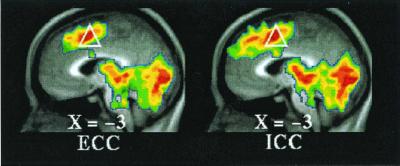
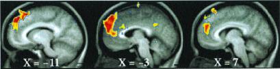
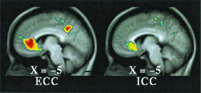
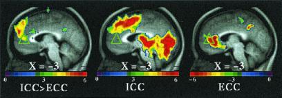
Medial prefrontal cortex (MPFC) is among those brain regions having the highest baseline metabolic activity at rest and one that exhibits decreases from this baseline across a wide variety of goal-directed behaviors in functional imaging studies. This high metabolic rate and this behavior suggest the existence of an organized mode of default brain function, elements of which may be either attenuated or enhanced. Extant data suggest that these MPFC regions may contribute to the neural instantiation of aspects of the multifaceted "self." We explore this important concept by targeting and manipulating elements of MPFC default state activity. In this functional magnetic resonance imaging (fMRI) study, subjects made two judgments, one self-referential, the other not, in response to affectively normed pictures: pleasant vs. unpleasant (an internally cued condition, ICC) and indoors vs. outdoors (an externally cued condition, ECC). The ICC was preferentially associated with activity increases along the dorsal MPFC. These increases were accompanied by decreases in both active task conditions in ventral MPFC. These results support the view that dorsal and ventral MPFC are differentially influenced by attentiondemanding tasks and explicitly self-referential tasks. The presence of self-referential mental activity appears to be associated with increases from the baseline in dorsal MPFC. Reductions in ventral MPFC occurred consistent with the fact that attention-demanding tasks attenuate emotional processing. We posit that both self-referential mental activity and emotional processing represent elements of the default state as represented by activity in MPFC. We suggest that a useful way to explore the neurobiology of the self is to explore the nature of default state activity.
Figures
References
-
- Shulman G L, Fiez J A, Corbetta M, Buckner R L, Miezin F M, Raichle M E, Petersen S E. J Cognit Neurosci. 1997;9:648–663. - PubMed
-
- Paus T, Koski L, Caramanos Z, Westbury C. NeuroReport. 1998;9:R37–R47. - PubMed
-
- Bush G, Luu P, Posner M I. Trends Cognit Sci. 2000;4:215–222. - PubMed
-
- Morris R, Pandya D N, Petrides M. J Comp Neurol. 1999;407:183–192. - PubMed
Publication types
MeSH terms
Grants and funding
LinkOut - more resources
Full Text Sources
Other Literature Sources
