. 2001 Mar 27;98(7):4125-9.
doi: 10.1073/pnas.071045598. Epub 2001 Mar 20.

Pectate lyase A, an enzymatic subunit of the Clostridium cellulovorans cellulosome

Affiliations

Pectate lyase A, an enzymatic subunit of the Clostridium cellulovorans cellulosome

Y Tamaru et al. Proc Natl Acad Sci U S A. .

Abstract

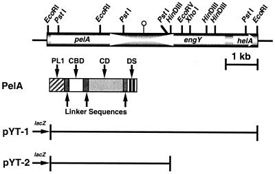
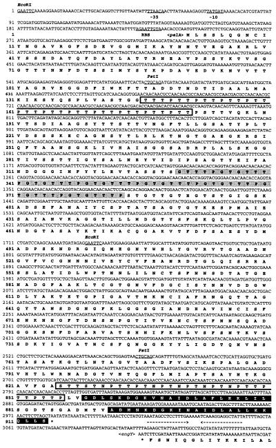
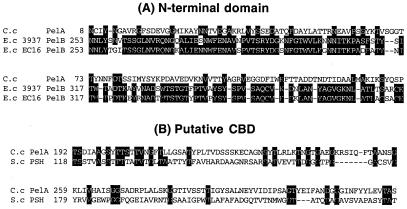
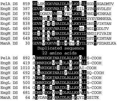
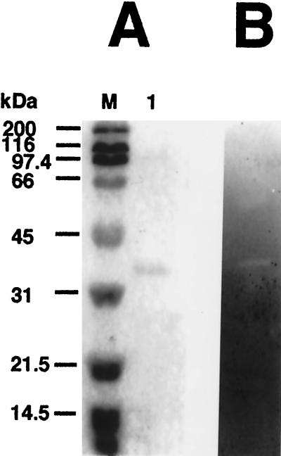
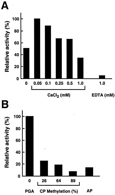
Clostridium cellulovorans uses not only cellulose but also xylan, mannan, pectin, and several other carbon sources for its growth and produces an extracellular multienzyme complex called the cellulosome, which is involved in plant cell wall degradation. Here we report a gene for a cellulosomal subunit, pectate lyase A (PelA), lying downstream of the engY gene, which codes for cellulosomal enzyme EngY. pelA is composed of an ORF of 2,742 bp and encodes a protein of 914 aa with a molecular weight of 94,458. The amino acid sequence derived from pelA revealed a multidomain structure, i.e., an N-terminal domain partially homologous to the C terminus of PelB of Erwinia chrysanthemi belonging to family 1 of pectate lyases, a putative cellulose-binding domain, a catalytic domain homologous to PelL and PelX of E. chrysanthemi that belongs to family 4 of pectate lyases, and a duplicated sequence (or dockerin) at the C terminus that is highly conserved in enzymatic subunits of the C. cellulovorans cellulosome. The recombinant truncated enzyme cleaved polygalacturonic acid to digalacturonic acid (G2) and trigalacturonic acid (G3) but did not act on G2 and G3. There have been no reports available to date on pectate lyase genes from Clostridia.


Figures

Figure 1
Figure 1
Restriction enzyme maps of pYT-1 and domain structure of the PelA polypeptide. Thin lines correspond to vector plasmid DNA. pYT-1 contains the entire 6.7-kb EcoRI fragment. The pectate lyase gene, pelA, was located on a 3.7-kb fragment designated as pYT-2. The white arrows indicate the coding sequences for the PelA, EngY, and HelA polypeptides, respectively. The different domains of PelA are named and filled with different patterns. PL1, pectate lyase family 1; CD, catalytic domain.
Figure 2
Figure 2
Nucleotide and deduced amino acid sequences of pelA. The putative promoter (−35 and −10) and ribosome-binding site sequences are underlined. Palindrome sequences are indicated by arrows facing each other. The stop codon is indicated by *. Proline-threonine- or asparagine-serine-threonine-rich regions are boxed. GTTTPGT repeats are shadowed. The DS (or dockerin) is highlighted.
Figure 3
Figure 3
Alignment of the N-terminal domain (A) and putative CBD domain (B) of PelA with those of other enzymes. Amino acids that are conserved in three sequences (A) and two sequences (B) are boxed. −, gaps left to improve alignment. Numbers refer to amino acid residues at the start of the respective lines; all sequences are numbered from Met-1 of the peptide. C.c, Clostridium cellulovorans; E.c, Erwinia chrysanthemi; S.c, Streptomyces coelicolor.
Figure 4
Figure 4
Alignment of pectate lyase family 4 catalytic domains of C. cellulovorans (C.c) PelA, Bacillus species (B.s) PelH, and E. chrysanthemi (E.c) PelL and PelX. Amino acids that are conserved in the four sequences are boxed. −, gaps left to improve alignment. Numbers refer to amino acid residues at the start of the respective lines; all sequences are numbered from Met-1 of the peptide.
Figure 5
Figure 5
Alignment of the DSs (or dockerins) of cellulosomal subunits of C. cellulovorans. Amino acids that are conserved in at least five of the nine sequences are highlighted. The numbers refer to amino acid residues at the start of the respective lanes; all sequences are numbered from Met-1 of the peptide.
Figure 6
Figure 6
Analysis of the purified rPelA by SDS/PAGE (A) and zymogram (B). Lane M, standard markers [myosin (200 kDa), β-galactosidase (116 kDa), phosphorylase B (97.4 kDa), BSA (66 kDa), ovalbumin (45 kDa), carbonic anhydrase (31 kDa), soybean trypsin inhibitor (21.5 kDa), and lysozyme (14.5 kDa)]; lane A 1, purified rPelA.
Figure 7
Figure 7
Enzymatic properties of PelA. (A) The influence of Ca2+ was tested by using various concentrations of CaCl2 in 50 mM Tris⋅HCl (pH 8) and 0.2% polygalacturonic acid. The cation requirement was confirmed by the addition of 1 mM EDTA. (B) The influence of the degree of pectin methylation on rPelA activity. Activity of rPelA on polygalacturonic acid (PGA), citrus pectins (CP) with a degree of methyl esterification from 26% to 89%, and apple pectin (AP) was determined by the standard assay condition.
Figure 8
Figure 8
TLC of the hydrolysis products of digalacturonic acid (G2), trigalacturonic acid (G3), and polygalacturonic acid (PG) with the purified rPelA. The reaction mixtures contained 50 mM Tris⋅HCl (pH 8), 0.05 mM CaCl2, 5 units of enzymatic activity/ml, and 1 mg of the substrates/ml. Incubations were performed at 37°C for 12 h after the addition of rPelA or without enzyme.

References

    1. Albersheim P, Killias U. Arch Biochem Biophys. 1962;97:107–115. - PubMed
    1. Barras F, van Gijsegem F, Chatterjee A K. Annu Rev Phytopathol. 1994;32:201–234.
    1. Collmer A, Keen N T. Annu Rev Phytopathol. 1986;24:383–409.
    1. Lietzke S E, Yoder M D, Keen N T, Jurnak F. Plant Physiol (Rockville) 1994;106:849–862. - PMC - PubMed
    1. Sleat R, Mah R A, Robinson R. Appl Environ Microbiol. 1984;48:88–93. - PMC - PubMed

Publication types

Associated data

LinkOut - more resources