. 2001 Mar 27;98(7):3808-13.
doi: 10.1073/pnas.240456398. Epub 2001 Mar 20.

Role of the cytoplasmic tyrosines of beta 1A integrins in transformation by v-src

Affiliations

Role of the cytoplasmic tyrosines of beta 1A integrins in transformation by v-src

T Sakai et al. Proc Natl Acad Sci U S A. .

Abstract

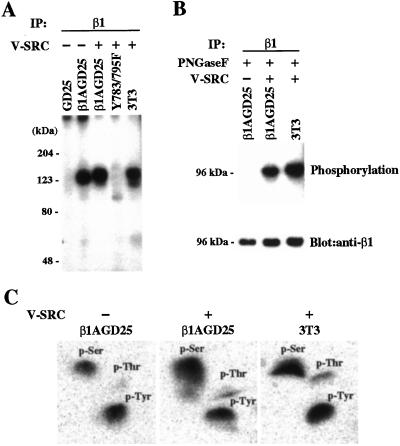
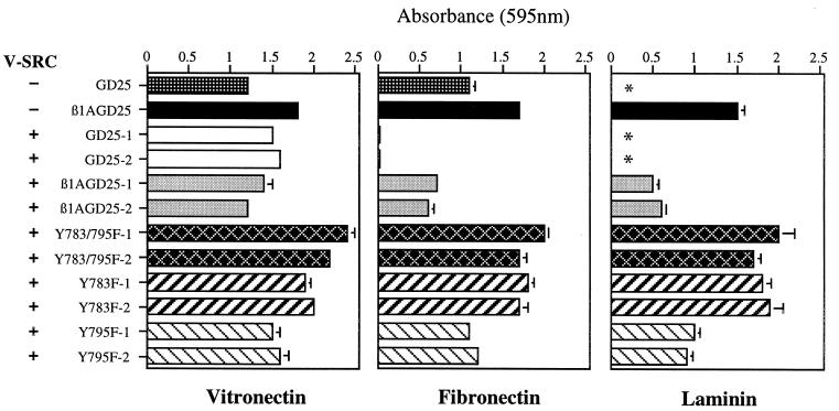
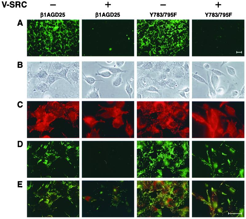
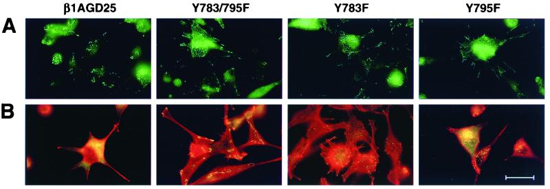
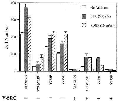
GD25 cells lacking beta 1 integrins or expressing beta 1A with mutations of conserved cytoplasmic tyrosines (Y783, Y795) to phenylalanine have poor directed migration to platelet-derived growth factor or lysophosphatidic acid when compared with GD25 cells expressing wild-type beta 1A. We studied the effects of v-src on these cells. Transformation with v-src caused tyrosine and serine phosphorylation of wild-type beta1 A but not of Y783/795F doubly mutated beta 1A. v-src-transformed cells had rounded and/or fusiform morphology and poor assembly of fibronectin matrix. Adhesion to fibronectin or laminin and coupling of focal contacts to actin-containing cytoskeleton were preserved in transformed Y783/795F cells but lost on transformation when beta 1A was wild type. Transformed Y783/795F cells also retained ability, albeit limited, to migrate across filters, whereas transformed cells with wild-type beta 1A were unable to transverse filters. Studies of single tyrosine mutants showed that the more important tyrosine for retaining ability to adhere, assemble focal contacts, and migrate is Y783. These results suggest that overactive phosphorylation of cytoplasmic residues of beta 1A, particularly Y783, accounts in part for the phenotype of v-src-transformed cells.


Figures

Figure 1
Figure 1
Phosphorylation of β1 integrin. (A) Autoradiogram of SDS/PAGE gel of [32P]orthophosphate-labeled proteins immunoprecipitated with monoclonal antibody MB1.2 to β1 from lysates of metabolically labeled nontransformed GD25 cells lacking β1A, nontransformed β1A-GD25 cells, v-src-transformed β1A-GD25 cells, v-src-transformed Y783/795F cells, and v-src-transformed 3T3 cells. Samples were reduced. The positions of the molecular mass markers are indicated (Left). (B) (Upper) Autoradiogram of poly(vinylidene difluoride) membrane of immunoprecipitates from lysates of nontransformed β1A-GD25 cells and v-src-transformed β1A-GD25 cells and 3T3 cells after PNGaseF treatment. (Lower) The same membrane immunoblotted with rabbit antibody against cytoplasmic domain of β1A. The positions of the molecular mass markers are indicated (Left). (C) Phosphoamino acid analysis of the bands of 123–128 kDa immunoprecipitated from nontransformed β1A-GD25 and v-src-transformed β1A-GD25 and 3T3 cells shown in A. Shown are phosphorimages of two-dimensional TLC plates. Positions of unlabeled phosphotyrosine, phosphoserine, and phosphothreonine, detected with ninhydrin, are indicated.
Figure 2
Figure 2
Attachment of v-src-transformed GD25 cells expressing no β1A, wild-type β1A, or mutant β1A on vitronectin, fibronectin, or laminin-1. Bars represent the mean of attachment activity quantified by spectrophotometric analysis at 595 nm after staining of adherent cells with bromophenol blue. Error bars represent ± SD of quadruplicate experiments. Absorbance because of nonspecific cell adhesion as measured on albumin-coated wells was less than 0.1 and has been subtracted. Two different clones in each v-src-transformed mutant (clones 1 and 2) were analyzed. GD25, β1-deficient cells; β1GD25, GD25 cells expressing wild-type B1A; other cells are designated by mutation(s). *, β1A-deficient GD25 cells had no detectable adherence to laminin-1.
Figure 3
Figure 3
Deposition of fibronectin in 3-day culture and colocalization of exogenous fibronectin and β1A in short-term assay in nontransformed and v-src-transformed β1A-GD25 or Y783/795F cells. (A) Immunofluorescence of fibronectin matrix after 3 days of culture in serum-containing medium. (B–E) Assembly of FITC-fibronectin during a 1-h period beginning 4 h after seeding of cells on surfaces coated with fibronectin. After fixation, staining for β1 integrin was performed by using antibody MB1.2 and lissamine rhodamine-labeled anti-rat IgG. The same field was photographed for cellular morphology by phase (B), β1A (C), FITC-fibronectin (D), and double fluorescence of β1A (red) and FITC-fibronectin (green) (E). (Bar = 35 μm.)
Figure 4
Figure 4
Visualization of focal contacts and associated structures in v-src-transformed β1A-GD25 or mutant β1A cells on fibronectin-coated substrata. (A) Single immunofluorescent detection of β1A. (B) Double immunofluorescent detection of vinculin (green) and rhodamine-phalloidin (red). β1A-GD25, GD25 cells expressing wild-type β1A; other cells are designated by mutation(s). Microscopic analysis of multiple cells in multiple fields indicated the β1- and vinculin-containing focal contacts were present in >40% of transformed Y783F and Y783/795F cells but in <5% of β1A cells and <20% of Y795F cells. (Bar = 30 μm.)
Figure 5
Figure 5
Cell migration through fibronectin-coated 5-μm filters in response to LPA or platelet-derived growth factor (PDGF). LPA (500 nM) or PDGF (10 ng/ml) was in the lower chamber. Each represents the mean of cell number per 0.16-mm2 field. Error bars indicate ± SD of quadruplicate determinations. β1A-GD25, GD25 cells expressing wild-type β1A; other cells are designated by mutation(s).

References

    1. Rous P. J Exp Med. 1911;13:397–411. - PMC - PubMed
    1. Jove R, Hanafusa H. Annu Rev Cell Biol. 1987;3:31–56. - PubMed
    1. Parsons J T, Weber M J. Curr Top Microbiol Immunol. 1989;147:79–127. - PubMed
    1. Burridge K, Fath K, Kelly T, Nuckolls G, Turner C E. Annu Rev Cell Biol. 1988;4:487–525. - PubMed
    1. Chambers A F, Tuck A B. Anticancer Res. 1988;8:861–872. - PubMed

Publication types