. 2001 Mar;44(3):608-17.
doi: 10.1002/1529-0131(200103)44:3<608::AID-ANR109>3.0.CO;2-2.

Cyclic tensile strain suppresses catabolic effects of interleukin-1beta in fibrochondrocytes from the temporomandibular joint

Affiliations

Cyclic tensile strain suppresses catabolic effects of interleukin-1beta in fibrochondrocytes from the temporomandibular joint

S Agarwal et al. Arthritis Rheum. 2001 Mar.

Abstract

Objective: To discern the effects of continuous passive motion on inflamed temporomandibular joints (TMJ).

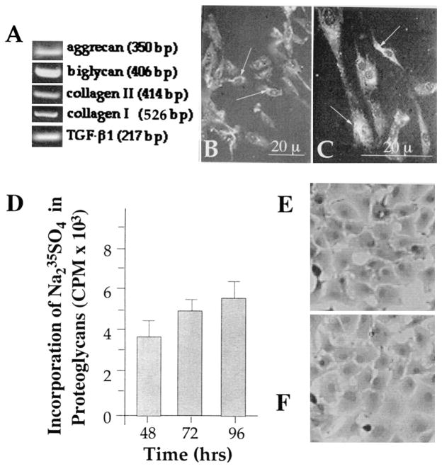
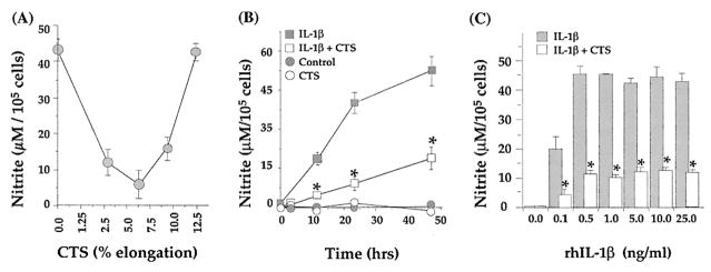
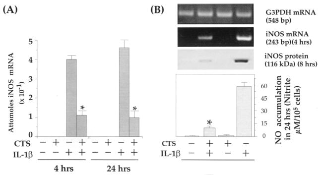
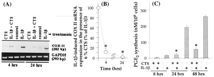
Methods: The effects of continuous passive motion on TMJ were simulated by exposing primary cultures of rabbit TMJ fibrochondrocyte monolayers to cyclic tensile strain (CTS) in the presence of recombinant human interleukin-1beta (rHuIL-1beta) in vitro. The messenger RNA (mRNA) induction of rHuIL-1beta response elements was examined by semiquantitative reverse transcriptase-polymerase chain reaction. The synthesis of nitric oxide was examined by Griess reaction, and the synthesis of prostaglandin E2 (PGE2) was examined by radioimmunoassay. The synthesis of proteins was examined by Western blot analysis of the cell extracts, and synthesis of proteoglycans via incorporation of 35S-sodium sulfate in the culture medium.

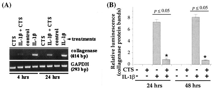
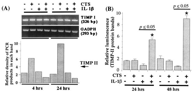
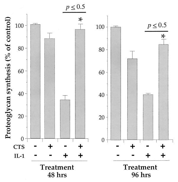
Results: Exposure of TMJ fibrochondrocytes to rHuIL-1beta resulted in the induction of inducible nitric oxide synthase (iNOS) and cyclooxygenase 2 (COX-2), which were paralleled by NO and PGE2 production. Additionally, IL-1beta induced significant levels of collagenase (matrix metalloproteinase 1 [MMP-1]) within 4 hours, and this was sustained over a period of 48 hours. Concomitant application of CTS abrogated the catabolic effects of IL-1beta on TMJ chondrocytes by inhibiting iNOS, COX-2, and MMP-1 mRNA production and NO, PGE2, and MMP-1 synthesis. CTS also counteracted cartilage degradation by augmenting expression of mRNA for tissue inhibitor of metalloproteinases 2 that is inhibited by rHuIL-1beta. In parallel, CTS also counteracted rHuIL-1beta-induced suppression of proteoglycan synthesis. Nevertheless, the presence of an inflammatory signal was a prerequisite for the observed CTS actions, because fibrochondrocytes, when exposed to CTS alone, did not exhibit any of the effects described above.

Conclusion: CTS acts as an effective antagonist of rHuIL-1beta by potentially diminishing its catabolic actions on TMJ fibrochondrocytes. Furthermore, CTS actions appear to involve disruption/regulation of signal transduction cascade of rHuIL-1beta upstream of mRNA transcription.


Figures

Figure 1
Figure 1
Phenotypic characteristics of rabbit temporomandibular joint (TMJ) fibrochondrocytes. A, Rabbit fibrochondrocytes exhibiting the presence of aggrecan, biglycan, type I collagen, type II collagen, and transforming growth factor β1 (TGF-β1)–specific mRNA, as assessed by reverse transcriptase–polymerase chain reaction. B and C. Rabbit TMJ disk chondrocytes showing the presence of aggrecan (B) and biglycan (C) by immunofluorescence (indicated by arrows). D, Presence of proteoglycans in TMJ disk chondrocytes as assessed by incorporation of Na235SO4 into chondroitin sulfate proteoglycans during the last 8 hours of the incubation. The proteoglycans were measured in a total of 2 × 10⁶ cells in triplicate as described in Materials and Methods. Bars represent the mean and SEM of triplicate values. E and F, Morphology of fibrochondrocytes without treatment (E) and following exposure to cyclic tensile strain for 24 hours (F).
Figure 2
Figure 2
A, Effect of various magnitudes of equibiaxial cyclic tensile strain (CTS) on recombinant human interleukin-1β (rhIL-1β)–induced production of nitric oxide (NO) in temporomandibular joint (TMJ) fibrochondrocytes. TMJ chondrocytes were incubated with rHuIL-1β (1 ng/ml) and simultaneously exposed to 0%, 3%, 6%, 9%, or 12% CTS for 24 hours. B, Time-dependent inhibition of rhIL-1β–induced production of NO by CTS. TMJ condylar chondrocytes were untreated, or were exposed to 6% CTS, to 1 ng/ml rhIL-1β, or to rhIL-1β and CTS for 4, 12, 24, or 48 hours. C, Effect of CTS on production of NO in response to various concentrations of rhIL-1β. Chondrocytes were treated with 0, 0,1, 0.5, 1, 5, 10, or 25 ng/ml of rHuIL-1β for 24 hours with or without simultaneous exposure to 6% CTS for 24 hours. Accumulation of NO in the culture supernatant was assessed by Griess reaction. Data represent the mean and SEM of triplicate values in all experiments. * = P < 0.05 as compared with cells treated with IL-1β alone.
Figure 3
Figure 3
CTS-mediated inhibition of recombinant human IL-1β (rHuIL-1β)–dependent inducible nitric oxide synthase (iNOS) expression and production of NO in TMJ fibrochondrocytes. A, Quantitative reverse transcriptase–polymerase chain reaction (RT-QCPCR) analysis of iNOS mRNA expression, showing inhibition of IL-1β (1 ng/ml)–induced mRNA production by CTS in TMJ fibrochondrocytes. B, Parallel experiments showing CTS-mediated inhibition of rHuIL-1β–induced iNOS mRNA synthesis, iNOS protein synthesis, and total accumulation of NO in culture supernatants of the TMJ fibrochondrocytes. Control cells and those treated with CTS alone did not exhibit iNOS gene products in RT-QCPCR, iNOS protein in Western blot analysis, or production of NO in culture supernatants. Results represent the mean and SEM in 1 of 3 separate experiments, showing similar data. * = P < 0.05 as compared with cells treated with IL-1β alone. See Figure 2 for other definitions.
Figure 4
Figure 4
Inhibition of recombinant human IL-1β (rHuIL-1β)–dependent cyclooxygenase 2 (COX-II) mRNA expression and prostaglandin E2 (PGE2) synthesis by CTS in TMJ fibrochondrocytes. A, COX-II mRNA expression in chondrocytes either untreated or subjected to 1 ng/ml rHuIL-1β, 6% CTS, or rHuIL-1β and CTS for 4 or 24 hours, showing a marked inhibition of IL-1β–dependent mRNA expression of COX-II. B, Inhibition of COX-II mRNA expression by 6% CTS in the presence of IL-1β as compared with cells treated with IL-1β alone. C, PGE2 synthesis in chondrocytes subjected to treatment regimens described in A for 8, 24, or 48 hours. PGE2 synthesis was measured in the culture supernatants by radioimmunoassay. Data in A represent 1 of 3 separate experiments. Data in B and C represent means and SEM of triplicate values. * = P < 0.05 as compared with cells treated with IL-1β alone. See Figure 2 for other definitions.
Figure 5
Figure 5
Effect of CTS on recombinant human IL-1β (rHuIL-1β)–dependent collagenase mRNA expression and synthesis in TMJ fibrochondrocytes. A, Expression of collagenase mRNA in chondrocytes either untreated or subjected to rHuIL-1β, CTS, or rHuIL-1β and CTS for 4 or 24 hours. B, Collagenase synthesis by fibrochondrocytes subjected to treatment regimens described in A for 24 or 48 hours. Collagenase synthesis was measured as the relative intensity of each band in Western blot analysis. Data in A represent 1 of 3 separate experiments. Data in B represent means and SEM of triplicate values. See Figure 2 for other definitions.
Figure 6
Figure 6
Effect of CTS on recombinant human IL-1β (rHuIL-1β)–dependent inhibition of tissue inhibitor of metalloproteinases (TIMP) mRNA expression. A, Expression of mRNA for TIMP I and TIMP II in TMJ chondrocytes either untreated or exposed to 1 ng/ml rHuIL-1β, 6% CTS, or rHuIL-1β and CTS. The mRNA expression was assessed by reverse transcriptase–polymerase chain reaction using 1 μg RNA from cells in each group. Amplification of GAPDH mRNA was used to ensure equal input in all lanes. B, TIMP II synthesis in cells subjected to treatment regimens described in A. Data in A represent 1 of 3 separate experiments. Data in B represent means and SEM of triplicate values. See Figure 2 for other definitions.
Figure 7
Figure 7
Effect of CTS on recombinant human IL-1β (rHuIL-1β)–dependent inhibition of proteoglycan synthesis in TMJ fibrochondrocytes. Chondrocytes were either untreated or exposed to 1 ng/ml rHuIL-1β, 6% CTS, or rHuIL-1β and CTS for 48 or 96 hours. Total proteoglycans were assessed by incorporation of Na2-35SO4 during the last 8 hours of incubation followed by extraction of glycosaminoglycans with 0.5M NaOH and separated on PD-10 size exclusion chromatography columns. Data represent means and SEM of triplicate values in 1 of 3 separate experiments. See Figure 2 for other definitions.

References

    1. Kopp S. Degenerative and inflammatory temporomandibular joint disorders: clinical perspectives. In: Sessle BJ, Bryant PS, Dionne RA, editors. Progress in pain research and management. Vol 4. Temporomandibular disorders and related pain conditions. Seattle (WA): ISAP Press; 1995. pp. 119–32.
    1. Albani S, Carson DA. Etiology and pathogenesis of rheumatoid arthritis. In: Koopman WJ, editor. Arthritis and allied health conditions: a textbook of rheumatology. Vol. 1. Baltimore (MD): Williams & Wilkins; 1997. pp. 979–91.
    1. Mahan PE, Alling CC. Temporomandibular joint anatomy, function, and pathofunction. In: Mahan PE, Alling CC III, editors. Facial pain. Philadelphia: Lea & Febiger; 1991. pp. 197–216.
    1. Zarb GA, Carlsson GE. Osteoarthrosis/osteoarthritis. In: Zarb GA, Carlsson GE, Sessle BJ, Mohl ND, editors. Temporomandibular joint and masticatory muscle disorders. Copenhagen: Munksgaard; 1994. pp. 298–327.
    1. Israel HA, Diamond BE, Saed-Nejad F, Ratcliffe A. Correlation between arthroscopic diagnosis of osteoarthritis and synovitis of the human temporomandibular joint and keratan sulfate levels in the synovial fluid. J Oral Maxillofac Surg. 1997;55:210–7. - PubMed

Publication types

MeSH terms