

The specific architecture of the anterior stroma accounts for maintenance of corneal curvature
- PMID: 11264134
- PMCID: PMC1723934
- DOI: 10.1136/bjo.85.4.437
The specific architecture of the anterior stroma accounts for maintenance of corneal curvature
Abstract
Aim: To analyse the human corneal stroma in extreme hydration to discover if its structure is responsible for corneal stability.
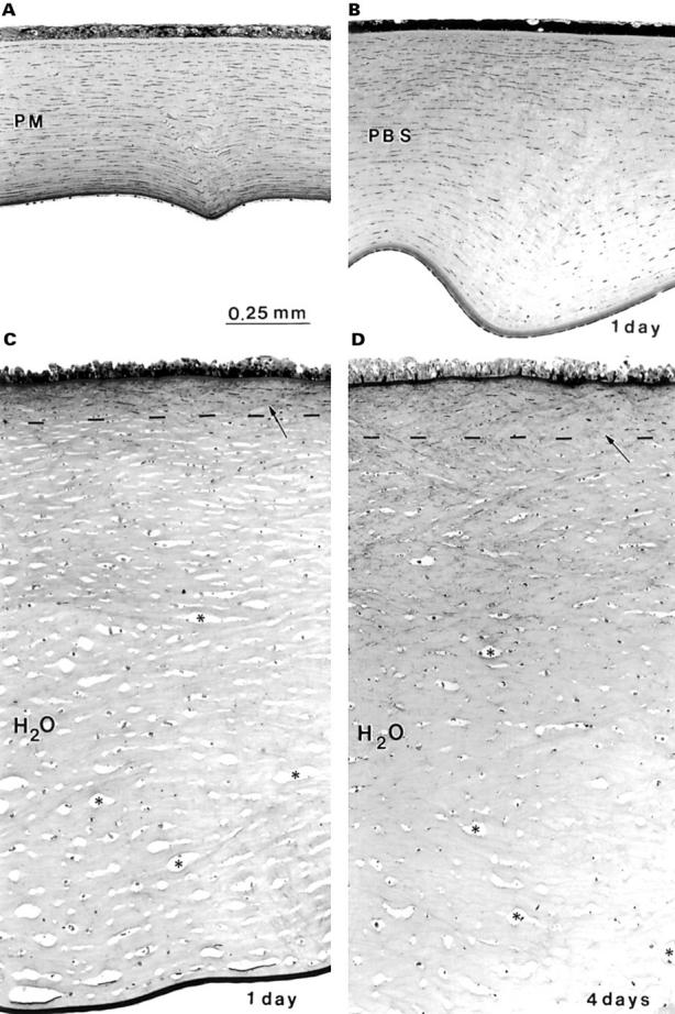
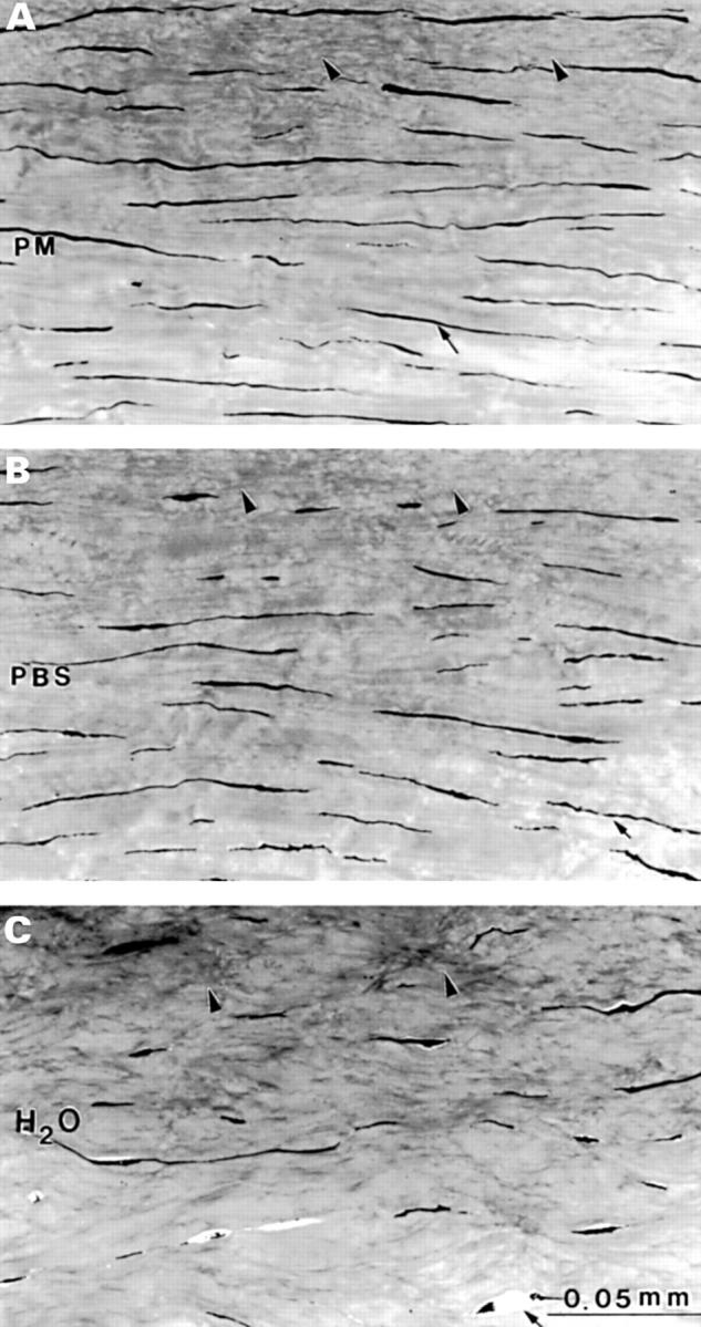
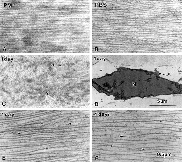
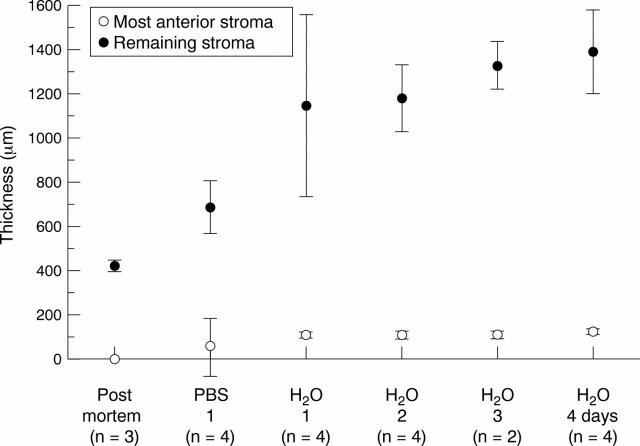
Methods: Corneas in several hydration states were used: postmortem control corneas (PM; n=3), corneas left for 1 day in phosphate buffered saline (PBS; n=4), and corneas left for 1 day (n=4), 2 days (n=4), 3 days (n=2), and 4 days (n=4) in deionised water. All corneas were fixed under standardised conditions and processed for light and electron microscopy. In addition, two fresh corneas from the operating theatre were studied which were processed 6 months after storage in sodium cacodylate buffer.
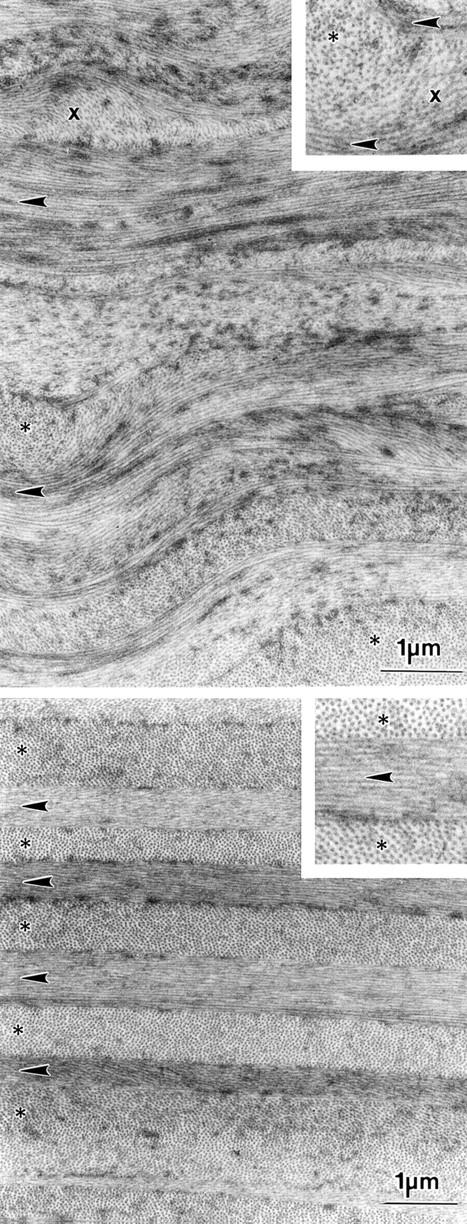
Results: After 1 day in deionised water maximal stromal swelling was reached which did not change up to 4 days. The stroma of deionised water corneas (1400 microm) was much thicker than that of PBS corneas (650 microm) and PM corneas (450 microm). Deionised water treatment led to disappearance of all keratocytes leaving only remnants of nuclei and large interlamellar spaces. In these specimens the distance between the collagen fibres had increased significantly, but the diameter of the collagen fibres did not seem to be affected. A remarkable observation was that the most anterior part of the stroma (100-120 microm) in all deionised water specimens and those stored for 6 months in buffer was not swollen, indicating that the tightly interwoven anterior lamellae are resistant to extreme non-physiological hydration states.
Conclusions: The rigidity of the most anterior part of the corneal stroma in extreme hydration states points to an important role in maintenance of corneal curvature. Since a large part of this rigid anterior part of the stroma is either removed (PRK) or intersected (LASIK), it is possible that in the long run patients who underwent refractive surgery may be confronted with optical problems.
Figures
Comment in
-
The architecture of the corneal stroma.Br J Ophthalmol. 2001 Apr;85(4):379-81. doi: 10.1136/bjo.85.4.379. Br J Ophthalmol. 2001. PMID: 11264120 Free PMC article. No abstract available.
References
MeSH terms
Substances
LinkOut - more resources
Full Text Sources
Other Literature Sources