

Intravitreal invading cells contribute to vitreal cytokine milieu in proliferative vitreoretinopathy
- PMID: 11264138
- PMCID: PMC1723908
- DOI: 10.1136/bjo.85.4.461
Intravitreal invading cells contribute to vitreal cytokine milieu in proliferative vitreoretinopathy
Abstract
Aim: To examine the contribution of infiltrating cells in the local production of cytokines within the vitreous of patients with proliferative vitreoretinopathy (PVR).
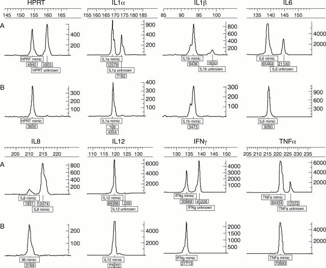
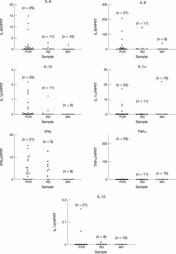
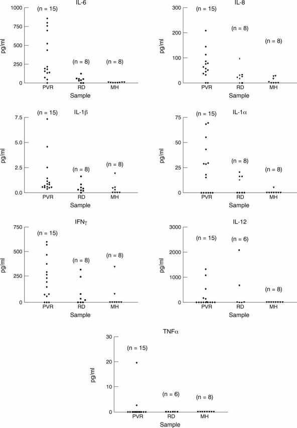
Methods: The presence of mRNA coding for IL-6, IL-8, IL-1beta, IL-1alpha, TNFalpha, IFNgamma, IL-12, and HPRT was investigated in 25 vitreous samples from patients with PVR, 11 vitreous samples from patients with retinal detachment (RD) not complicated by PVR, and 10 vitreous samples from patients with macular hole (MH). A quantitative reverse transcriptase polymerase chain reaction (RT-PCR) using an internal competitor was used to investigate these samples. From these samples, 15 PVR, 8 RD, and 8 MH were analysed for the protein levels of the same cytokines using enzyme linked immunosorbent assay (ELISA). Spearman correlation was used to test any association between mRNA and cytokine protein levels, as an indicator of the contribution these cells make to the intravitreal cytokine milieu.
Results: A strong correlation was found between mRNA and their respective cytokine levels (protein products) for IL-6, IL-8, IL-1beta, IL-1alpha, TNFalpha, IFNgamma (Spearman r = 0.83, 0.73, 0.67, 0.91, 0.73, and 0.73 respectively), but not for IL-12. The median levels of IL-6, IL-8, IL-1beta, and IFNgamma mRNA and their respective cytokines were significantly higher (p <0.05) in patients with PVR than in those with macular hole. There was no statistically significant difference in the median levels of IL-1alpha mRNA between PVR and MH but the cytokine IL-1alpha was detected at a significantly higher level in PVR compared with MH patients. Between PVR and RD patients, there was no statistically significant difference in mRNA levels for all the investigated cytokines (p >0.05) except for IL-6 where there was a statistical significance (p= 0.038). In contrast, the median levels of IL-6, IL-8, and IL-1beta cytokines were significantly higher (p <0.05) in patients with PVR than in those with RD, whereas for IL-1alpha and IFNgamma no significant statistical difference was detected between PVR and RD patients (p >0.05). When results of RD and MH patients were compared, a statistical difference was only detected in mRNA levels of INFgamma (p = 0.008). However, no difference was detected for INFgamma (protein product) or for any of the other cytokines between RD and MH patients.
Conclusion: Levels of both protein and mRNA encoding IL-6, IL-8, IL-1beta, and IFNgamma is significantly increased in vitreous samples from patients with PVR. The strong correlation between ELISA detectable cytokines (protein products) and their respective mRNA levels suggest that intravitreal, invasive cells are the major source of these cytokines, with the exception of IL-12. Cells invading the vitreous do not appear to locally produce IL-12 mRNA. This would appear to implicate cells peripheral to the vitreal mass as the major source of this cytokine.
Figures
References
Publication types
MeSH terms
Substances
LinkOut - more resources
Full Text Sources
Other Literature Sources
Research Materials
Miscellaneous