. 2001 Apr;132(7):1531-41.
doi: 10.1038/sj.bjp.0703976.

Mitogenic effect of oxidized low-density lipoprotein on vascular smooth muscle cells mediated by activation of Ras/Raf/MEK/MAPK pathway

Affiliations

Mitogenic effect of oxidized low-density lipoprotein on vascular smooth muscle cells mediated by activation of Ras/Raf/MEK/MAPK pathway

C M Yang et al. Br J Pharmacol. 2001 Apr.

Abstract

1. It has been demonstrated that oxidized low-density lipoprotein (OX-LDL) is a risk factor in atherosclerosis by stimulating vascular smooth muscle cell (VSMC) proliferation. However, the mechanisms of OX-LDL-induced cell proliferation are not completely understood. Therefore, we investigated the effect of OX-LDL on cell proliferation associated with mitogen-activated protein kinase (MAPK) activation in rat cultured VSMCs. 2. Both native-LDL (N-LDL) and OX-LDL induced a time- and concentration-dependent incorporation of [(3)H]-thymidine in VSMCs. 3. OX-LDL induced time- and concentration-dependent phosphorylation of p42/p44 MAPK. Pretreatment of these cells with pertussis toxin or U73122 attenuated the OX-LDL-induced responses. 4. Pretreatment with PMA for 24 h, preincubation with a PKC inhibitor staurosporine or the tyrosine kinase inhibitors, genistein and herbimycin A for 1 h, substantially reduced [(3)H]-thymidine incorporation and p42/p44 MAPK phosphorylation induced by OX-LDL. 5. Removal of Ca(2+) by BAPTA/AM or depletion of the internal Ca(2+) pool by thapsigargin significantly inhibited OX-LDL-induced [(3)H]-thymidine incorporation and p42/p44 MAPK phosphorylation. 6. OX-LDL-induced [(3)H]-thymidine incorporation and p42/p44 MAPK phosphorylation was inhibited by PD98059 (an inhibitor of MEK1/2) and SB203580 (an inhibitor of p38 MAPK) in a concentration-dependent manner. 7. Overexpression of dominant negative mutants of Ras (H-Ras-15A) and Raf (Raf-N4) significantly suppressed MEK1/2 and p42/p44 MAPK activation induced by OX-LDL and PDGF-BB, indicating that Ras and Raf may be required for activation of these kinases. 8. These results suggest that the mitogenic effect of OX-LDL is mediated through a PTX-sensitive G protein-coupled receptor that involves the activation of the Ras/Raf/MEK/MAPK pathway similar to that of PDGF-BB in rat cultured VSMCs.


Figures
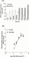
Figure 1
Figure 1
[3H]-thymidine incorporation induced by N-LDL or OX-LDL in VSMCs. (A) Time course, after 24 h in serum-free medium, the cells were stimulated with vehicle, N-LDL or OX-LDL at a concentration of 100 μg ml−1. The cells were labelled with 1 μCi ml−1 [3H]-thymidine for the times indicated in the continuous presence of N-LDL or OX-LDL. (B) Concentration-dependence, the cells were stimulated with various concentrations of N-LDL or OX-LDL. After stimulation for 8 h, VSMCs were labelled with 1 μCi ml−1 [3H]-thymidine for another 16 h in the continued presence of N-LDL or OX-LDL. The incorporation of [3H]-thymidine was determined as described in Methods. Data are expressed as the mean±s.e.mean of three separate experiments determined in triplicate.
Figure 2
Figure 2
Time course of OX-LDL-stimulated p42/p44 MAPK phosphorylation in VSMCs. The cells were grown to confluency, made quiescent by serum-deprivation for 24 h and incubated with 100 μg ml−1 OX-LDL for 2 – 60 min. The cell lysates were subjected to 10% SDS – PAGE and transferred to nitrocellulose membrane. Western blot analysis was performed using an antiserum reactive with anti-phospho-p42/p44 MAPK polyclonal antibody for activated p42/p44 MAPK and anti-p42 MAPK antibody for the total p42 MAPK as an indicator of protein loading in each well. Bands were visualized by an ECL method. Data are expressed as the mean±s.e.mean of three separate experiments. *P<0.01, compared with the respective basal level.
Figure 3
Figure 3
Concentration-dependence of OX-LDL-stimulated p42/p44 MAPK phosphorylation in VSMCs. The cells were grown to confluency, made quiescent by serum-deprivation for 24 h and incubated with various concentrations of OX-LDL for 5 min. The cell lysates were subjected to 10% SDS – PAGE and transferred to nitrocellulose membrane. Bands were visualized by an ECL method as described in Figure 2. Data are expressed as the mean±s.e.mean of three separate experiments. *P<0.01, compared with the respective basal level.
Figure 4
Figure 4
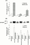
Effects of pertussis toxin and U73122 on DNA synthesis and p42/p44 MAPK phosphorylation induced by OX-LDL in VSMCs. (A) The cells were grown to confluency, made quiescent by serum-deprivation and preincubated with pertussis toxin (PTX, 100 ng ml−1) for 24 h or U73122 (10 μM) for 1 h, and then stimulated with vehicle or 100 μg ml−1 OX-LDL. After 8 h incubation, cells were labelled with 1 μCi ml−1 [3H]-thymidine for another 16 h in the continuous presence of OX-LDL. The incorporation of [3H]-thymidine was determined as described in Methods. Data are expressed as the mean±s.e.mean of three separate experiments determined in triplicate. *P<0.01, compared with the cells exposed to OX-LDL. (B) For MAPK experiment, after incubation with PTX for 24 h or U73122 for 1 h, the cells were stimulated with 100 μg ml−1 OX-LDL for 5 min. The cell lysates were subjected to 10% SDS – PAGE and transferred to nitrocellulose membrane. Bands were visualized by an ECL method as described in Figure 2. Data are expressed as the mean±s.e.mean of three separate experiments. *P<0.01, compared with the cells exposed to OX-LDL.
Figure 5
Figure 5
Involvement of PKC in DNA synthesis and p42/p44 MAPK phosphorylation induced by OX-LDL in VSMCs. (A) The cells were grown to confluence, made quiescent by serum-deprivation for 24 h. The cells were preincubated with 1 μM staurosporine (STA) for 1 h, or 1 μM PMA for 24 h before stimulated with vehicle or 100 μg ml−1 OX-LDL. The incorporation of [3H]-thymidine was determined as described in Figure 4. Data are expressed as the mean±s.e.mean of three separate experiments determined in triplicate. *P<0.01, compared with the cells exposed to OX-LDL. (B) For MAPK experiment, after incubation with STA for 1 h, or PMA for 24 h, the cells were stimulated with 100 μg ml−1 OX-LDL for 5 min. The cell lysates were subjected to 10% SDS – PAGE and transferred to nitrocellulose membrane. Bands were visualized by an ECL method as described in Figure 2. Data are expressed as the mean±s.e.mean of three separate experiments. *P<0.05, compared with the cells exposed to OX-LDL.
Figure 6
Figure 6
Effect of BAPTA plus EGTA on OX-LDL-induced [3H]-thymidine incorporation and p42/p44 MAPKphosphorylation in VSMCs. (A) The cells were grown to confluence, made quiescent by serum-deprivation for 24 h. The cells were preincubated with 30 μM BAPTA and 5 mM EGTA for 1 h and then stimulated with vehicle or 100 μg ml−1 OX-LDL. The incorporation of [3H]-thymidine was determined as described in Figure 4. Data are expressed as the mean±s.e.mean of three separate experiments determined in triplicate. *P<0.01, compared with the cells exposed to OX-LDL. (B) For MAPK experiment, after incubation with BAPTA and EGTA for 1 h, the cells were stimulated with 100 μg ml−1 OX-LDL for 5 min. The cell lysates were subjected to 10% SDS – PAGE and transferred to nitrocellulose membrane. Bands were visualized by an ECL method as described in Figure 2. Data are expressed as the mean±s.e.mean of three separate experiments. *P<0.01, compared with the cells exposed to OX-LDL.
Figure 7
Figure 7
Effects of diltiazem, ryanodine, and thapsigargin on OX-LDL-induced [3H]-thymidine incorporation in VSMCs. The cells were grown to confluence, made quiescent by serum-deprivation for 24 h. The cells were preincubated with 10 μM diltiazem (Dil), ryanodine or thapsigargin (TG) for 1 h and then stimulated with vehicle or 100 μg ml−1 OX-LDL. The incorporation of [3H]-thymidine was determined as described in Figure 4. Data are expressed as the mean±s.e.mean of three separate experiments determined in triplicate. *P<0.001, compared with the cells exposed to OX-LDL.
Figure 8
Figure 8
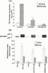
Involvement of tyrosine kinase in DNA synthesis and p42/p44 MAPK phosphorylation induced by OX-LDL in VSMCs. (A) The cells were grown to confluence, made quiescent by serum-deprivation for 24 h. The cells were preincubated with 10 μM genistein and 10 μM herbimycin A for 1 h, and then stimulated with vehicle or 100 μg ml−1 OX-LDL. The incorporation of [3H]-thymidine was determined as described in Figure 4. Data are expressed as the mean±s.e.mean of three separate experiments determined in triplicate. *P<0.01, compared with the cells exposed to OX-LDL. (B) For MAPK experiment, after incubation with genistein (GEN) and herbimycin A (HER) for 1 h, the cells were stimulated with 100 μg ml−1 OX-LDL for 5 min. The cell lysates were subjected to 10% SDS – PAGE and transferred to nitrocellulose membrane. Bands were visualized by an ECL method as described in Figure 2. Similar results were obtained in three independent experiments.
Figure 9
Figure 9
Effects of MAPK kinase inhibitors on OX-LDL-stimulated DNA synthesis and p42/p44 MAPK phosphorylation in VSMCs. (A) The cells were grown to confluence, made quiescent by serum-deprivation for 24 h. The cells were preincubated with 10 μM PD98059 and 10 μM SB203580 for 1 h, and then stimulated with vehicle or 100 μg ml−1 OX-LDL. The incorporation of [3H]-thymidine was determined as described in Figure 4. Data are expressed as the mean±s.e.mean of three separate experiments determined in triplicate. *P<0.01, compared with the cells exposed to OX-LDL. (B) For MAPK experiment, after incubation with increasing concentrations of PD98059 and SB203580 for 1 h, the cells were stimulated with 100 μg ml−1 OX-LDL for 5 min. The cell lysates were subjected to 10% SDS – PAGE and transferred to nitrocellulose membrane. Bands were visualized by an ECL method as described in Figure 2. Data are expressed as the mean±s.e.mean of three separate experiments. *P<0.01, compared with the cells exposed to OX-LDL.
Figure 10
Figure 10
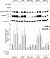
Requirement of Ras and Raf for OX-LDL- and PDGF-BB-induced activation of MEK1/2 and p42/p44 MAPK in VSMCs. Cells were transfected with plasmids encoding pZIP-NeoSV, H-Ras-15A, pCGN, or Raf-N4, and then stimulated with OX-LDL (100 μg ml−1) or PDGF-BB (20 ng ml−1) for 10 min. The cell lysates were subjected to 10% SDS – PAGE and transferred to nitrocellulose membrane. Western blot analysis was performed using an antiserum reactive with the anti-phospho-MEK1/2, anti-phospho-p42/p44 MAPK, and total p42 MAPK (as a control) polyclonal antibody. Bands were visualized by an ECL method. *P<0.05; #P<0.001, compared with respective control.
Figure 11
Figure 11
Effects of BQ-123 and losartan on OX-LDL-stimulated p42/p44 MAPK phosphorylation in VSMCs. The cells were grown to confluence, made quiescent by serum-deprivation for 24 h. The cells were preincubated with 10 μM BQ-123 or 10 μM losartan for 1 h, and then stimulated with vehicle or 100 μg ml−1 OX-LDL for 5 min. The cell lysates were subjected to 10% SDS – PAGE and transferred to nitrocellulose membrane. Bands were visualized by an ECL method as described in Figure 2. Data are expressed as the mean±s.e.m. of three separate experiments.

Similar articles

Cited by

References

    1. ABDELLATIF M., PACKE S.E., MICHAEL L.H., ZHANG D., CHARNG M.J., SCHNEIDER M.D. A ras-dependent pathway regulates RNA polymerase II phosphorylation in cardiac myocytes: implications for cardiac hypertrophy. Mol. Cell. Biol. 1998;18:6729–6736. - PMC - PubMed
    1. AUGE N., PIERAGGI M.-T., THIERS J.-C., NEGRE-SALVAYRE A. Proliferative and cytotoxic effects of mildly oxidized low density lipoproteins on vascular smooth muscle cells. Biochem. J. 1995;309:1015–1020. - PMC - PubMed
    1. AUTIO I., JAAKOLA O., SOLAKIVI T., NIKKARI T. Oxidized low-density lipoprotein is chemotactic for arterial smooth muscle cells in culture. FEBS Lett. 1990;277:247–249. - PubMed
    1. BLENIS J. Signal transduction via the MAP kinases: Proceed at your own RSK. Proc. Natl. Acad. Sci. U.S.A. 1993;90:5889–5892. - PMC - PubMed
    1. BOULANGER C.M., TANNER F.C., B'EA M.L., HAHN A.W., WERNER A., LUSCHER T.F. Oxidized low density lipoproteins induce mRNA expression and release of endothelin from human and porcine endothelium. Cir. Res. 1992;70:1191–1197. - PubMed

Publication types

MeSH terms

Substances