. 2001 Apr 1;21(7):2288-97.
doi: 10.1523/JNEUROSCI.21-07-02288.2001.

Changes in microtubule stability and density in myelin-deficient shiverer mouse CNS axons

Collaborators, Affiliations

Changes in microtubule stability and density in myelin-deficient shiverer mouse CNS axons

L L Kirkpatrick et al. J Neurosci. .

Abstract

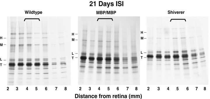
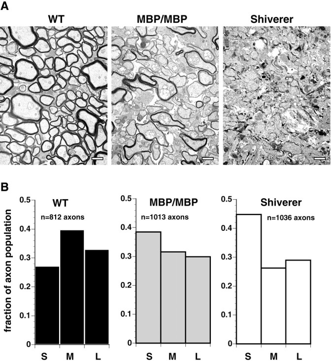
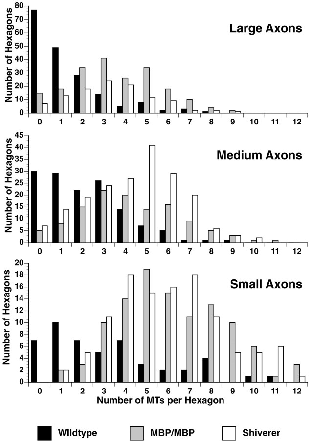
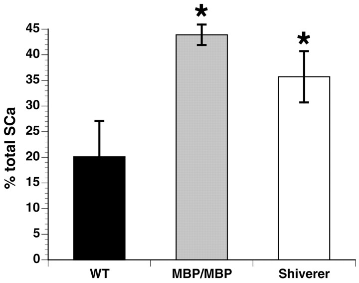
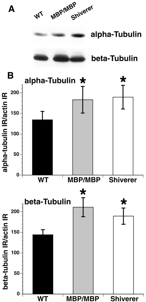
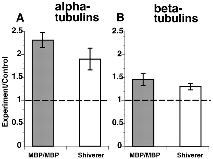
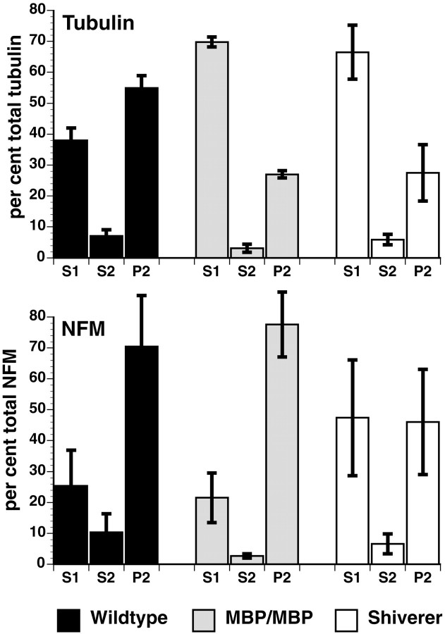
Altered axon-Schwann cell interactions in PNS myelin-deficient Trembler mice result in changed axonal transport rates, neurofilament and microtubule-associated protein phosphorylation, neurofilament density, and microtubule stability. To determine whether PNS and CNS myelination have equivalent effects on axons, neurofilaments, and microtubules in CNS, myelin-deficient shiverer axons were examined. The genetic defect in shiverer is a deletion in the myelin basic protein (MBP) gene, an essential component of CNS myelin. As a result, shiverer mice have little or no compact CNS myelin. Slow axonal transport rates in shiverer CNS axons were significantly increased, in contrast to the slowing in demyelinated PNS nerves. Even more striking were substantial changes in the composition and properties of microtubules in shiverer CNS axons. The density of axonal microtubules is increased, reflecting increased expression of tubulin in shiverer, and the stability of microtubules is drastically reduced in shiverer axons. Shiverer transgenic mice with two copies of a wild-type myelin basic protein transgene have an intermediate level of compact myelin, making it possible to determine whether the actual level of compact myelin is an important regulator of axonal microtubules. Both increased microtubule density and reduced microtubule stability were still observed in transgenic mouse nerves, indicating that signals beyond synaptogenesis and the mere presence of compact myelin are required for normal regulation of the axonal microtubule cytoskeleton.


Figures

Fig. 1.
Fig. 1.
Axonal transport of tubulins in shiverer, transgenic, and wild-type mouse optic nerve. Slow axonal transport rates were examined by segmental analysis.35S-methionine was injected into the vitreous of the mouse eye, and 21 d after injection, optic nerve–optic tracts were harvested and cut into 1 mm segments. Radioactively labeled proteins in each segment were resolved on SDS-PAGE gels, processed for fluorography, and exposed to film. Fluorographs of wild-type, MBP/MBP transgenic, and shiverer optic nerve show a wave of radioactively labeled SCa proteins traveling down the optic nerve–optic tract. Radiolabeled tubulin (T) and the neurofilament triplet subunits (H, M, and L) are distributed as a wave along the nerve in all three animals at 21 d after injection, but the position of the peak differs. The peak for tubulins is in axon segments 4–5 mm for wild-type and MBP/MBP nerves but in segments 5–6 mm for shiverer. Note that the fraction of SCa-labeled proteins in the tubulin band is higher in shiverer and MBP/MBP nerves than in wild type.
Fig. 2.
Fig. 2.
Distribution of axonal calibers is affected by myelination. A, Electron micrographs of optic nerve from wild-type (WT), transgenic (MBP/MBP), and shiverer (Shiverer) mice exhibit striking differences in myelination. No compact myelin is apparent in shiverer nerves, and compact myelin in MBP/MBP nerve is much reduced relative to wild type. B, Morphometric analysis of axon caliber in optic nerve from wild-type and mutant mice shows a shift in axon caliber with increasing levels of compact myelin. Optic nerve axons are among the smallest myelinated axons and are relatively uniform in size. However, 45% of the axons in shiverer nerve have an area of <0.2 mm2 [small (S)], in contrast to wild-type nerve in which only 27% fell into the S category. Most axons in wild-type nerve were in the medium (M; 40%) or large (L; 33%) categories, whereas in shiverer there were 26% in the M and 28% in the Lcategories. In MBP/MBP nerves, the axon area phenotype was intermediate, consistent with an intermediate level of compact myelin. As with shiverer nerve, the largest number of axons were categorized asS, but the fraction in S was only 38%, with 32% in M and 30% in L.
Fig. 3.
Fig. 3.
Morphometric analysis of MT distribution shows different densities in different caliber axons. The density of microtubules in wild type, MBP/MBP, and shiverer for different sized axons was analyzed as described previously (de Waegh et al., 1992) by determining the number of cytoskeletal elements in random hexagons. Size categories are as described in Figure 2 and Table 2. Morphometric analyses were conducted by overlaying a hexagonal grid over electron micrographs of the optic axonal cross sections printed at a final magnification of 140,000×. Each hexagon represented an area of 0.035 μm2. The number of microtubules per hexagon was scored, binned, and plotted. Microtubule densities are shifted to higher values in both shiverer and MBP/MBP axons for all sizes of axon, but the density was greatest in small axons (means of 6.0–6.1 per hexagon in shiverer and MBP/MBP; mean of 3.3 per hexagon in wild type) and correspondingly reduced in medium (4.2–4.4 vs 2.2 per hexagon) and large (3.3–3.5 vs 1.3 per hexagon) axons (Table 2). In all cases, the shiverer and MBP/MBP axons showed similar MT density distributions. These values may be converted to MTs per micrometer2, giving values for shiverer and MBP/MBP of 170 MTs/μm2 (S), 123 MTs/μm2 (M), and 97 MTs/μm2(L), as compared with 94 MTs/μm2 (S), 63 MTs/μm2 (M), and 37 MTs/μm2(L) for wild-type axons.
Fig. 4.
Fig. 4.
Tubulin levels may be quantitated from axonal transport studies. Differences in the amount of tubulin in optic axons may be seen by measuring the total amount of labeled tubulin in the nerve at a given time point and expressing it as a ratio of the total amount of labeled protein in the nerve at the same time. Tubulin represented 20% of labeled protein in wild-type axons, but was 44% of labeled protein in MBP/MBP axons and 36% in shiverer axons. This indicates that the amount of tubulin committed to slow axonal transport is increased in the absence of a normal complement of compact myelin.Asterisk indicates significant difference when compared with wild-type (p ≤ 0.02).
Fig. 5.
Fig. 5.
Total brain tubulin is increased relative to total brain actin in shiverer and MBP/MBP mice. A, Quantitative immunoblots using antibodies specific for α- and β-tubulin show that total brain tubulin immunoreactivity is increased in both shiverer and MBP/MBP mice. B, Normalization of tubulin immunoreactivity to actin immunoreactivity in the same samples allows a quantitative comparison of changes in tubulin immunoreactivity in brain. Brain α-tubulin immunoreactivity is increased by 36% in MBP/MBP brain and by 41% in shiverer brain relative to that seen in wild-type brains. Similarly, brain β-tubulin immunoreactivity is increased by 46% in MBP/MBP brain and by 31% in shiverer brain relative to that seen in wild-type brains. Asteriskindicates significant difference when compared with wild-type (p ≤ 0.01).
Fig. 6.
Fig. 6.
Quantitative Northern blots indicate that tubulin mRNA levels are increased in shiverer and MBP/MBP mouse brains. RNA fractions from retina were probed with oligonucleotides shared with all known mouse α- or β-tubulins as well as one for GAPDH as a loading control. After correction for mRNA load with GAPDH, a ratio of mutant to wild-type expression levels for both α- and β-tubulin was calculated. The levels of both α- and β-tubulin mRNA expression were significantly increased for shiverer and MBP/MBP relative to wild type. As with changes in tubulin protein levels, α- and β-tubulin RNA levels were significantly increased in MBP/MBP and shiverer mice relative to wild type (p = 0.0002 and 0.009 for α-tubulin, p = 0.02 and 0.007 for β-tubulin, in transgenic and shiverer, respectively).
Fig. 7.
Fig. 7.
Cold-calcium fractionation of SCa-labeled cytoskeletal proteins indicates that myelination affects the stability of the axonal cytoskeleton. Using our standard cold-calcium fractionation protocols (Kirkpatrick and Brady, 1994), the stability of the microtubule and neurofilament cytoskeletons may be analyzed. As in Trembler peripheral nerve, the amount of tubulin in the P2 fraction (top panel) is substantially reduced in the absence of myelin (shiverer). As with other parameters associated with the axonal microtubule cytoskeleton, the thin myelin sheath in MBP/MBP optic axons is not sufficient to increase the amount of cold-calcium-insoluble tubulin in P2 (top panel). In both mutant mice, cold-calcium-insoluble tubulin fractions are reduced by half, and extractable tubulin is correspondingly increased. Unlike Trembler peripheral nerve, however, a difference in the stability of the neurofilament cytoskeleton was also observed (bottom panel). When compact myelin was completely absent (shiverer), the amount of NFM found in the stable fraction dropped from 71 to 47% with a corresponding increase in the extractable form. However, even a thin myelin sheath was sufficient to reverse this effect, because MBP/MBP values are comparable with wild type. Similar changes in neurofilament stability were seen for NFL and NFH as well (Table 4). These data suggest that myelination leads to stabilization of both microtubule and neurofilament axonal cytoskeletons, but the effects on microtubules and neurofilaments are modulated independently.

Similar articles

Cited by

References

    1. Aguayo A, David S, Bray G. Influence of the glial environment on the elongation of axons after injury: transplantation studies in adult rodents. J Exp Biol. 1981;95:231–240. - PubMed
    1. Benley M, Aguayo A. Extensive elongation of axons from rat brain into peripheral nerve graft. Nature. 1982;296:150–152. - PubMed
    1. Brady S. Axonal transport methods and applications. In: Boulton A, Baker G, editors. Neuromethods, general neurochemical techniques. Humana; Clifton, NJ: 1985. pp. 419–476.
    1. Brady ST. Increases in cold-insoluble tubulin during aging. Soc Neurosci Abstr. 1984;10:273.
    1. Brady ST. Cytotypic specializations of the neuronal cytoskeleton and cytomatrix: implications for neuronal growth and regeneration. In: Haber B, Gorio A, Vellis JD, Perez-Polo JR, editors. Cellular and molecular aspects of neural development and regeneration. Springer; New York: 1988. pp. 311–322.

Publication types