. 2001 Apr 1;21(7):2298-307.
doi: 10.1523/JNEUROSCI.21-07-02298.2001.

A depletable pool of adenosine in area CA1 of the rat hippocampus

Affiliations

A depletable pool of adenosine in area CA1 of the rat hippocampus

T Pearson et al. J Neurosci. .

Abstract

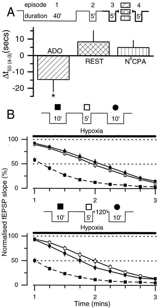
Adenosine plays a major modulatory and neuroprotective role in the mammalian CNS. During cerebral metabolic stress, such as hypoxia or ischemia, the increase in extracellular adenosine inhibits excitatory synaptic transmission onto vulnerable neurons via presynaptic adenosine A(1) receptors, thereby reducing the activation of postsynaptic glutamate receptors. Using a combination of extracellular and whole-cell recordings in the CA1 region of hippocampal slices from 12- to 24-d-old rats, we have found that this protective depression of synaptic transmission weakens with repeated exposure to hypoxia, thereby allowing potentially damaging excitation to both persist for longer during oxygen deprivation and recover more rapidly on reoxygenation. This phenomenon is unlikely to involve A(1) receptor desensitization or impaired nucleoside transport. Instead, by using the selective A(1) antagonist 8-cyclopentyl-1,3-dipropylxanthine and a novel adenosine sensor, we demonstrate that adenosine production is reduced with repeated episodes of hypoxia. Furthermore, this adenosine depletion can be reversed at least partially either by the application of exogenous adenosine, but not by a stable A(1) agonist, N(6)-cyclopentyladenosine, or by endogenous means by prolonged (2 hr) recovery between hypoxic episodes. Given the vital neuroprotective role of adenosine, these findings suggest that depletion of adenosine may underlie the increased neuronal vulnerability to repetitive or secondary hypoxia/ischemia in cerebrovascular disease and head injury.


Figures

Fig. 1.
Fig. 1.
Role of adenosine A1 receptors in the hypoxic depression of excitatory synaptic transmission. Pooled data, normalized to the prehypoxic fEPSP slope, for control (●;n = 24) and 200 nm DPCPX-treated (⋄;n = 5) slices showing the effect of a single 10 min hypoxic episode (denoted by the black bar).Inset shows typical fEPSPs taken at the time points indicated, before (a, d), during (b, e), and after (c,f) the hypoxic episode: control (a–c); 200 nm DPCPX (d–f). Note the differences in the fEPSPs at time points (b) and after (e) 3 min of hypoxia reflecting the A1 receptor-dependence of the hypoxic depression of the fEPSP. Calibration: 10 msec, 0.4 mV.
Fig. 2.
Fig. 2.
Exposure to hypoxia results in reduced sensitivity of synaptic transmission to subsequent hypoxia.A, A typical experiment in which two sequential hypoxic episodes were given to the same slice. A(1) shows fEPSP slope versus time. Labeled at time points a throughf are fEPSPs (inset above, stimulus artifacts are truncated) before (a, d), during (b, e), and after (c, f) two sequential hypoxic episodes (10 and 5 min, respectively). A(2), Superimposition of normalized data from the first 4 min of each hypoxic episode (▪, first episode; ○, second episode). Notice the resistance (conditioning) of the fEPSP to hypoxia during the second episode. The superimposed fEPSPs (b, e), both taken 2 min into the hypoxic episode, highlight this apparent acquired resistance to the effects of hypoxia. Calibration: 10 msec, 0.4 mV. B, Conditioning depends on the duration of the first hypoxic episode. Insets show pooled normalized data of the influence of the duration of the first hypoxic episodes (▪) of (from left to right) 2.25 (n = 11), 10 (n = 24), and 40 min (n = 21) on the decay of the fEPSP during the second hypoxic episode (○). Graph shows dependence of conditioning, expressed as the difference between the time to 50% depression of the fEPSP of the first and second episodes (Δt50: 2.25 min, n = 11; 5 min, n = 32; 10 min, n = 94; 20 min, n = 10; 40 min, n = 27) on the duration of initial hypoxic episode. Linethrough points follows the equation given in Results and gives a time constant of conditioning of 725 sec.
Fig. 3.
Fig. 3.
Role of adenosine A1 receptors in the hypoxic depression of excitatory synaptic transmission under whole-cell voltage-clamp conditions. Pooled data, normalized to the prehypoxic EPSC amplitude, for control (▪; n = 70) and 200 nm DPCPX-treated (⋄; n = 14) slices show the effect of a single 10 min hypoxic episode (denoted by theblack bar). Inset shows typical EPSCs taken at the time points indicated, before (a,d), during (b, e), and after (c, f) the hypoxic episode: control (a–c); 200 nm DPCPX (d–f). Note the differences in the fEPSPs at time points b and e after 10 min of hypoxia reflecting the A1 receptor-dependence of the hypoxic depression of the EPSC. Calibration: 40 msec, 50 pA.
Fig. 4.
Fig. 4.
Reduced sensitivity of whole-cell voltage-clamp EPSCs to hypoxia. A, Effect of two 10 min periods of hypoxia on EPSC during an individual experiment. Note reduced rate of depression of the EPSC during the second hypoxic episode (○) compared with the first (▪). Inset shows EPSCs taken at the times indicated. Calibration: 30 msec, 50 pA. B, Pooled data from 20 cells in which cells were exposed to two sequential 10 min periods of hypoxia (first episode, ▪; second episode, ○). Note increased resistance to the effects of hypoxia during the second hypoxic episode.
Fig. 5.
Fig. 5.
Conditioning depends on a reduction in extracellular adenosine and not changes in adenosine transport or A1 receptor desensitization. A, Incubation with the adenosine transport inhibitors NBTI (1 μm) and DIPY (5 μm), denoted by bar, greatly depressed the fEPSP (n = 8). When the depression had stabilized, two sequential 5 min hypoxic episodes (each denoted by a bar) were administered. This resulted in a complete depression of synaptic transmission and conditioning of the fEPSP that was no different (p = 0.82; unpairedt test) from that in the absence of NBTI/DIPY (inset histogram; NBTI/DIPY, n = 8; control, n = 32). Application of DPCPX (200 nm; n = 5), denoted bybar, confirmed the NBTI/DIPY-induced depression as being dependent on A1 receptors. Break in time course plot reflects ∼30 min. B, Pooled normalized data of fEPSP depressions to two sequential 10 min applications of 100 μm adenosine on the fEPSP (Δt50 = 1 ± 3 sec;n = 6). C, Pooled normalized data of fEPSP depressions to hypoxia while in the presence of 10 nmDPCPX (n = 7; ▪, first hypoxic episode; ■, second hypoxic episode) and for comparison controls (n = 24; ●, first hypoxic episode; ○, second hypoxic episode). For clarity, the shaded bars represent the Δt50 values for the control experiments (left) and DPCPX experiments (right). Inset is ahistogram comparing the magnitude of conditioning in control (n = 94) and 10 nmDPCPX-treated slices (n = 7). DPCPX-treated slices exhibited significantly greater conditioning (Student'st test, p < 0.0001), indicating a reduction in extracellular adenosine as the basis of conditioning.
Fig. 6.
Fig. 6.
Direct measurement of reduced adenosine release during repeated hypoxia. A, Output from the adenosine sensor during two sequential 5 min periods of hypoxia (black bar and between upward deflections of chart event marker). Note reduced adenosine release during second hypoxic episode. Calibration: 2 μm adenosine, 3 min. Break in chart record reflects ∼17 min. B, Field EPSPs taken at the times indicated inC. Calibration: 10 msec, 0.25 mV. C, Time course of hypoxic depression of fEPSP showing slower rate of depression (c vs d), similar maximal depression (e vs f), and more rapid recovery of transmission (g vs h) during the second hypoxic episode (○) compared with the first (▪).
Fig. 7.
Fig. 7.
Adenosine depletion in nominally Ca2+-free aCSF. Two sequential 5 min periods of hypoxia (black bars and upward deflections of chart event markers) separated by 30 min in a slice incubated in nominally Ca2+-free aCSF (2 mmCa2+ replaced by 2 mmMg2+) for >3 hr. Note large decrease in adenosine release in response to the second hypoxic episode. Calibration: 5 μm, 5 min.
Fig. 8.
Fig. 8.
Depletion of adenosine production during successive hypoxic episodes. Pooled data from three different experimental protocols showing the minute-by-minute profile of adenosine release during the first (▪) and second (○) hypoxic episodes (denoted by black bar). A, Two sequential 5 min hypoxic episodes in 2 mm extracellular Ca2+ (n = 8); B, two sequential 10 min hypoxic episodes in 2 mmextracellular Ca2+ (n = 8); C, two sequential 5 min hypoxic episodes in nominally Ca2+-free aCSF (2 mmCa2+ replaced with 2 mmMg2+; n = 6).
Fig. 9.
Fig. 9.
Replenishment of the depleted adenosine.A, Exogenous adenosine replenishes the depleted adenosine. After one 40 and two 5 min hypoxic episodes, slices were exposed to 20 μm adenosine (ADO;n = 10) or 30 nmN6CPA (N6CPA;n = 9) or allowed to rest for an equivalent time (15 min; REST; n = 6). A fourth 5 min hypoxic episode was then administered (inset: experimental protocol). A comparison was then made between thet50 values of the third and fourth hypoxia-induced depressions of the fEPSP (Δt50(4–3); in bold in protocol). The bar chart shows that the application of adenosine, but not of the selective A1 agonist N6CPA or an equivalent rest period, results in an acceleration of the rate of depression of the fEPSP by hypoxia (p < 0.05, unpaired t test).B, Replenishment by endogenous adenosine. Experiment in which a 10 (▪; dashed line) and 5 (■) min hypoxic episode was followed by a second 10 (●) min episode after a short interval (∼10 min; n = 13; top) or after a 2 hr interval (120′; n = 13;bottom). With a short rest, hypoxia causes further conditioning (rightward shift) between the second (■) and third (●) hypoxic episodes. However, a prolonged rest period of 2 hr between the second (■) and third (●) hypoxic episodes causes the third exposure to hypoxia to induce a more rapid depression of the fEPSP (leftward shift) throughout the hypoxic episode. These data argue for a replenishment of the depleted adenosine and against gradual deterioration in slice viability.

Similar articles

Cited by

References

    1. Arlinghaus L, Lee KS. Endogenous adenosine mediates the sustained inhibition of excitatory synaptic transmission during moderate hypoxia. Brain Res. 1996;724:265–268. - PubMed
    1. Berne RM, Rubio R, Curran GH. Release of adenosine from ischemic brain. Circ Res. 1974;35:262–271.
    1. Blumbergs PC. Pathology. In: Reilly P, Bullock R, editors. Head injury: pathophysiology and management of severe closed injury. Chapman & Hall; London: 1997. pp. 40–70.
    1. Chen J, Simon R. Ischemic tolerance in the brain. Neurology. 1997;48:306–311. - PubMed
    1. Cherubini E, Ben-Ari Y, Krnjevic K. Anoxia produces smaller changes in synaptic transmission, membrane potential, and input resistance in immature rat hippocampus. J Neurophysiol. 1989;62:882–895. - PubMed

Publication types

LinkOut - more resources