. 2001 Apr 1;21(7):2425-33.
doi: 10.1523/JNEUROSCI.21-07-02425.2001.

Agonist-induced internalization and trafficking of cannabinoid CB1 receptors in hippocampal neurons

Affiliations

Agonist-induced internalization and trafficking of cannabinoid CB1 receptors in hippocampal neurons

A A Coutts et al. J Neurosci. .

Abstract

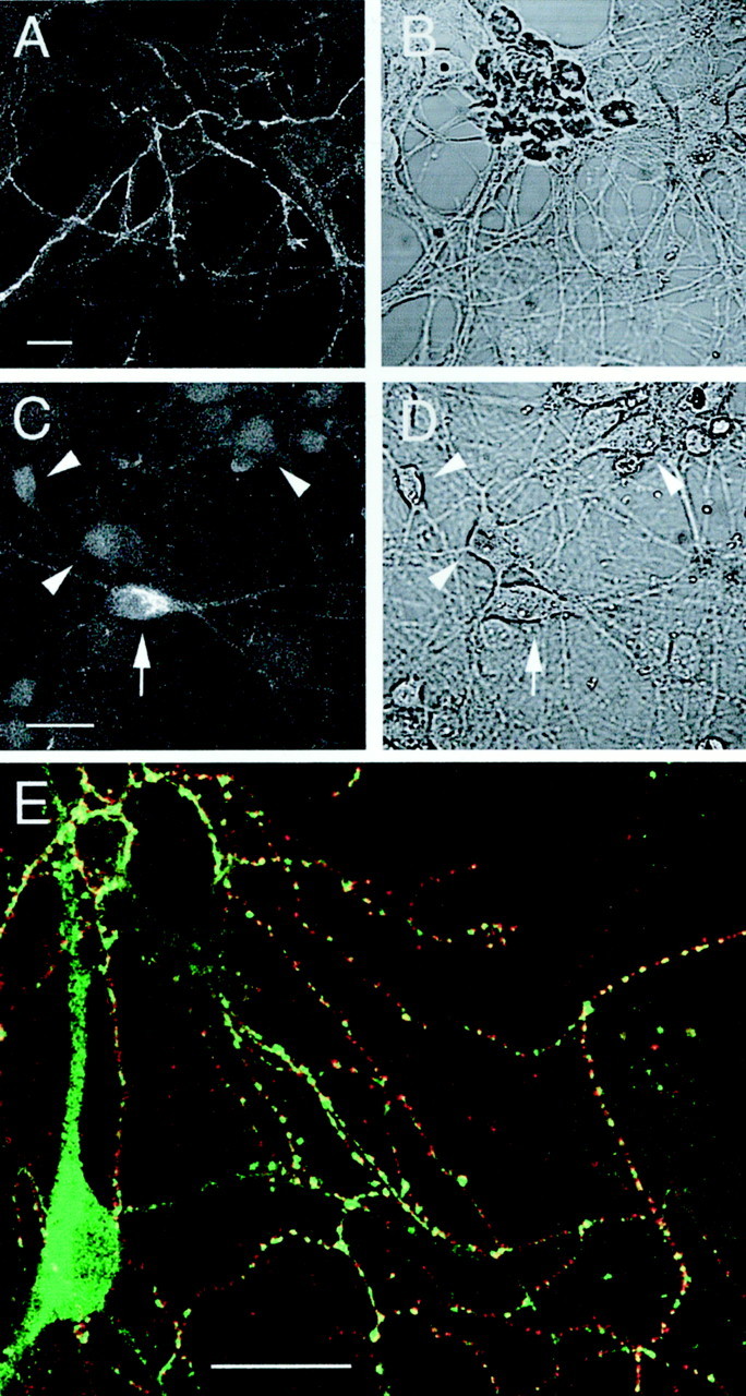
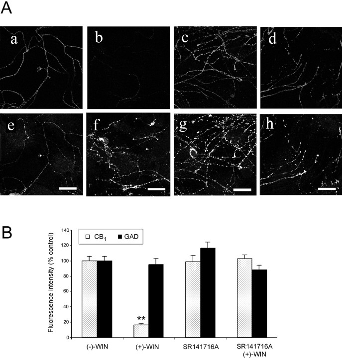
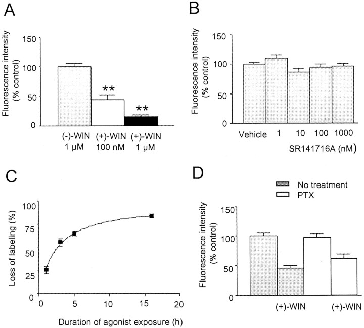
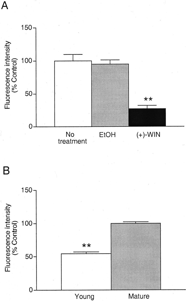
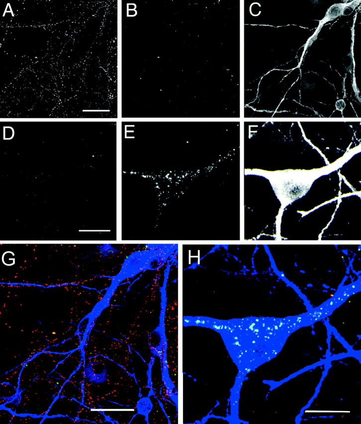
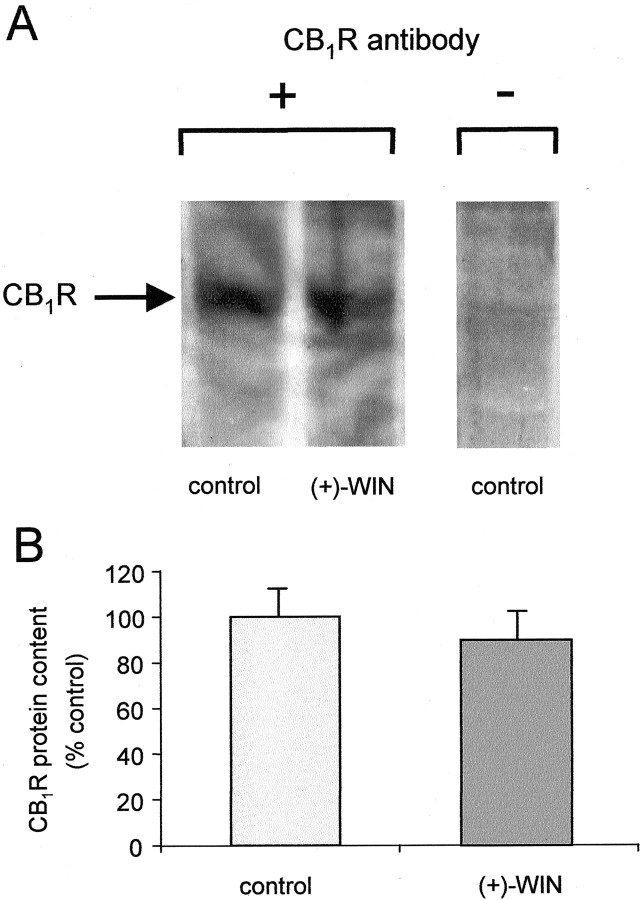
Agonist-induced internalization of G-protein-coupled receptors is an important mechanism for regulating receptor abundance and availability at the plasma membrane. In this study we have used immunolabeling techniques and confocal microscopy to investigate agonist-induced internalization and trafficking of CB(1) receptors in rat cultured hippocampal neurons. The levels of cell surface CB(1) receptor immunoreactivity associated with presynaptic GABAergic terminals decreased markedly (by up to 84%) after exposure to the cannabinoid agonist (+)-WIN55212, in a concentration-dependent (0.1-1 microm) and stereoselective manner. Inhibition was maximal at 16 hr and abolished in the presence of SR141716A, a selective CB(1) receptor antagonist. Methanandamide (an analog of an endogenous cannabinoid, anandamide) also reduced cell surface labeling (by 43% at 1 microm). Differential labeling of cell surface and intracellular pools of receptor demonstrated that the reduction in cell surface immunoreactivity reflects agonist-induced internalization and suggests that the internalized CB(1) receptors are translocated toward the soma. The internalization process did not require activated G-protein alpha(i) or alpha(o) subunits. A different pattern of cell surface CB(1) receptor expression was observed using an undifferentiated F-11 cell line, which had pronounced somatic labeling. In these cells substantial CB(1) receptor internalization was also observed after exposure to (+)-WIN55212 (1 microm) for relatively short periods (30 min) of agonist exposure. In summary, this dynamic modulation of CB(1) receptor expression may play an important role in the development of cannabinoid tolerance in the CNS. Agonist-induced internalization at presynaptic terminals has important implications for the modulatory effects of G-protein-coupled receptors on neurotransmitter release.


Figures

Fig. 1.
Fig. 1.
CB1 receptor immunoreactivity in cultured hippocampal neurons. Representative images depicting cell surface labeling of intact cells showing immunoreactivity associated with a network of fine fibers (A) and total labeling after fixation and permeabilization (C).B and D are corresponding bright-field images. Note the strong intracellular immunoreactivity associated with a neuronal somata (arrow). Neurons lacking somatic labeling are also indicated (arrowheads). Immunofluorescence images A and C arez projections of a series of confocal sections taken at 1–2 μm intervals. E shows merged, single plane confocal images from a dual-labeling experiment investigating the relationship between cell surface CB1 receptor clusters and inhibitory terminals, labeled with a monoclonal GAD antibody after permeabilization. Red corresponds to CB1receptor label (Cy3), green to GAD label (Alexa 488), and yellow to regions of overlap. Note the marked correspondence between CB1 receptor label and clusters of GAD immunoreactivity. Scale bars, 20 μm.
Fig. 2.
Fig. 2.
The effects of cannabinoid pretreatment on cell surface CB1 receptor labeling. A, Immunolabeling of cells pretreated for 16 hr with (−)-WIN55212 [(−)-WIN; a, e], (+)-WIN55212[(+)-WIN; b, f], (+)-WIN55212 with SR141716A (c, g), and SR141716A alone (d, h), all at 1 μm. Representative confocal images (single section) of CB1 receptor (a–d) and corresponding GAD (e–h) immunolabeling are depicted for each treatment. Scale bars, 20 μm.B, Quantitative histogram showing the effects of cannabinoid pretreatment on CB1 receptor labeling. Values are mean ± SEM of normalized relative to control fluorescence intensities obtained with (−)-WIN55212. For each paradigm 27 fibers from a minimum of three independent experiments were analyzed (**p < 0.01). Note that (+)-WIN55212 markedly inhibited the labeling of CB1 receptor immunoreactivity in the absence, but not in the presence, of SR141716A. GAD immunolabeling was unaffected by cannabinoid pretreatment.
Fig. 3.
Fig. 3.
Further characterization of the effects of cannabinoids on cell surface CB1 receptor expression.A, Histogram showing the inhibition of CB1receptor immunofluorescence on cells preincubated for 16 hr with (+)-WIN55212 (100 nm and 1 μm) compared with control cells preincubated with (−)-WIN55212 (1 μm).B, Histogram showing the mean fluorescence intensity of fibers after preincubation with SR141716A (1–1000 nm) compared with vehicle (control). C, Graph showing the effects of incubation time in (+)-WIN55212 (1 μm) on cell surface labeling. The mean level of fluorescence at each time interval was compared with that of cells treated with vehicle (control).D, Histogram showing the effects of overnight pretreatment of hippocampal cells with PTX (100 ng/ml) on the agonist-induced loss of cell surface labeling (expressed relative to vehicle controls). Neither the level of CB1 receptor expression nor the loss of cell surface labeling caused by treatment with (+)-WIN55212 (1 μm; 16 hr) were significantly affected by PTX (p > 0.05). Values are mean ± SEM; ** p < 0.01.
Fig. 4.
Fig. 4.
Actions of cannabinoids on immature hippocampal neurons. A, Histogram showing the inhibition of cell surface CB1 receptor immunofluorescence on fibers after exposure to (+)-WIN55212 (1 μm) or the equivalent concentration of vehicle (EtOH) for 72 hr immediately after plating and compared with untreated (control) cells. B, Histogram showing the relative level of CB1 receptor immunoreactivity expressed in young cultures (2 d) compared with control values obtained with mature neurons (9–14 d). Values are mean ± SEM (**p < 0.01).
Fig. 5.
Fig. 5.
Visualizing cell surface and internalized CB1 receptors in cultured hippocampal neurons using primary antibody prelabeling. Corresponding images from triple-labeling experiments comparing vehicle (A–C) with (+)-WIN55212 (1 μm; D–F) on cell surface and intracellular CB1 receptor immunofluorescence.A and D show surface CB1receptors; B and E show internalized receptors. In C and F the cell soma and proximal dendrites have been labeled with MAP-2 antibody. Images arez projections of a series of 11 confocal sections taken at 2 μm intervals. G and H show the corresponding merged color images for vehicle and (+)-WIN55212 treatment. Red corresponds to cell surface label (Cy3), green to internalized receptor (Alexa 488), and blue to MAP-2 (Cy5). Note how cell surface CB1 receptor-positive fibers are intertwined with and track the MAP-2-positive dendrites. The figure shows a representative experiment from seven determinations with similar findings, and in three of these, cells were subsequently colabeled with MAP-2. Scale bars: A, G, 25 μm; D, H, 15 μm.
Fig. 6.
Fig. 6.
Localization of cell surface and internalized CB1 receptors in F-11 cells. A–D, Confocal images of F-11 cells that had been incubated with vehicle alone (A, B) were compared with cells that had been treated with (+)-WIN55212 (1 μm; C, D). Cells were labeled for surface CB1 receptors (A, C) and internalized receptors after cell permeabilization (B, D). Note the loss of cell surface labeling and the appearance of internalized receptors after exposure to (+)-WIN55212. Scale bars, 50 μm.
Fig. 7.
Fig. 7.
Western analysis of CB1 receptor immunoreactivity in rat cultured hippocampal cells. Cells were pretreated with (+)-WIN55212 or vehicle (control) at 37°C for 16 hr.A, The membrane proteins were immunostained with (+) or without (−) exposure to CB1 receptor primary antibody before secondary antisera. In both treatments a specific band of 61 kDa (CB1R) was detected. This band was not markedly altered by (+)-WIN55212 treatment. B, Densitometric analysis of the 61 kDa band (CB1R) from Western blots of rat cultured hippocampal cells as described in (A). Mean ± SEM; p > 0.05; n = 3.

References

    1. Bernard V, Laribi O, Levey AI, Bloch B. Subcellular redistribution of m2 muscarinic acetylcholine receptors in striatal interneurons in vivo after acute cholinergic stimulation. J Neurosci. 1998;18:10207–10218. - PMC - PubMed
    1. Bouaboula M, Perrachon S, Milligan L, Canat X, Rinaldi-Carmona M, Portier M, Barth F, Calandra B, Pecceu F, Lupker J, Maffrand J-P, Le Fur G, Casellas P. A selective inverse agonist for central cannabinoid receptor inhibits mitogen-activated protein kinase activation stimulated by insulin or insulin-like growth factor 1. Evidence for a new model of receptor/ligand interactions. J Biol Chem. 1997;272:22330–22339. - PubMed
    1. Coutts AA, Pertwee RG. Inhibition by cannabinoid receptor agonists of acetylcholine release from the guinea-pig myenteric plexus. Br J Pharmacol. 1997;121:1557–1566. - PMC - PubMed
    1. Coutts AA, Brewster N, Ingram T, Razdan RK, Pertwee RG. Comparison of novel cannabinoid partial agonists and SR141716A in the guinea-pig small intestine. Br J Pharmacol. 2000;129:645–652. - PMC - PubMed
    1. Devane WA, Hanus L, Breuer A, Pertwee RG, Stevenson LA, Griffin G, Gibson D, Mandelbaum A, Etinger A, Mechoulam R. Isolation and structure of a brain constituent that binds to the cannabinoid receptor. Science. 1992;258:1946–1949. - PubMed

Publication types

Substances

LinkOut - more resources