. 2001 Apr 1;21(7):2462-73.
doi: 10.1523/JNEUROSCI.21-07-02462.2001.

Coincidence detection or temporal integration? What the neurons in somatosensory cortex are doing

Affiliations

Coincidence detection or temporal integration? What the neurons in somatosensory cortex are doing

S A Roy et al. J Neurosci. .

Abstract

To assess the impact of thalamic synchronization on cortical responsiveness, we used conditional cross-correlation analysis to measure the probability of neuronal discharges in somatosensory cortex as a function of the time between discharges in pairs of simultaneously recorded neurons in the ventrobasal thalamus. Among 26 neuronal trios, we found that thalamocortical efficacy after synchronous thalamic activity was nearly twice as large as the efficacy rate obtained when pairs of thalamic neurons discharged asynchronously. Nearly half of these neuronal trios displayed cooperative effects in which the cortical discharge probability after synchronous thalamic events was larger than could be predicted from the efficacy rate of individual thalamic discharges. In these cases of heterosynaptic cooperativity, thalamocortical efficacy declined to asymptotic levels when the interspike intervals were >6-8 msec. These results indicate that thalamic synchronization has a significant impact on cortical responsiveness and suggest that neuronal synchronization may play a critical role in the transmission of sensory information from one brain region to another.


Figures

Fig. 1.
Fig. 1.
Representative experiment illustrating relationship between thalamic synchronization and thalamocortical coordination (experiment TC12). A, Receptive fields for neurons in the ventrobasal complex (vb1, vb2) and secondary somatosensory cortex (SII). Red circlesindicate airjet stimulation sites. B, PSTHs of neuronal responses to 200 trials of airjet stimulation. Binwidths, 25 msec.C, Raw CCGs displaying significant amounts of correlated activity as indicated by peaks exceeding the 95% confidence limits (red lines). Each CCG was based on the sum of discharges occurring in response to all stimulus configurations. Binwidths, 1 msec. D, Template indicating how patterns of thalamic and cortical activity are displayed in the snowflake histograms.Dashed lines represent time axes for displaying interspike intervals for each pair of neurons (e.g., TSII− Tvb1). Red lines represent synchronous time axes for displaying instances in which neurons discharge at the same time (e.g., Tvb1 = Tvb2);points along the horizontal red line indicate instances of thalamic synchronization. Blue lines depict concentric hexagons that represent 5 msec intervals along each interspike interval axis. As shown by the spike trains and their corresponding points in the template, synchronized thalamic events and subsequent cortical discharges appear to the right of the central origin. E, Summed snowflake histogram for experiment TC12 based on discharges occurring in response to all stimulus configurations. White bins on the horizontal time axis (red arrow) reflect a large number of cortical discharges that occurred immediately after the thalamic neurons discharged simultaneously. Thalamocortical interactions for each thalamic neuron are indicated byfaint diagonal bands (blue arrows). Total stimulus-induced discharges and a color-coded legend appear at theleft. Binwidths, 0.5 msec.
Fig. 2.
Fig. 2.
Cortical responses to varying amount of thalamic synchronization. Top panel, Summed snowflake histograms, illustrated as in Figure 1, displaying coordinated activity for three separate experiments (TC4, TC15, andTC16). Binwidths, 0.5 msec. Bottom panel, Raw CCGs indicating the proportion of thalamic activity that was synchronized in these experiments. The correlation coefficients appear next to the CCG peak. Binwidths, 1.0 msec.
Fig. 3.
Fig. 3.
Effects of different stimuli and their combined administration on the coordination of thalamic and cortical activity in experiment TC15. A, Receptive fields and airjet stimulation sites. B, PSTHs illustrating neuronal responses in the ventrobasal thalamus and SII cortex during 200 stimulation trials.C, Snowflake histograms illustrating how thalamocortical coordination varied in response to different airjets or their combination. All panels illustrated as in Figure 1.
Fig. 4.
Fig. 4.
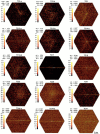
Variations in the pattern of thalamocortical coordination recorded across different neuronal trios. Each snowflake histogram represents the summed response to all stimulus configurations as shown in Figures 1 and 2. Some recording experiments are represented by more than one snowflake histogram because multiple neurons were often recorded from the two thalamic electrodes (e.g., TC5) or from the electrodes in SII cortex (e.g., TC13, TC14, andTC44).
Fig. 5.
Fig. 5.
Limitations of snowflake histograms for depicting thalamocortical interactions. A and B illustrate two sets of spike trains that cannot be plotted in a snowflake histogram because only two of the three neurons discharged within the analysis interval (±15 msec). C and D illustrate two sets of spike trains that are depicted by an identical pair of points in the snowflake histogram shown to the right. Hence, identical data points in a snowflake histogram may represent two isolated instances of heterosynaptic integration (C) or an instance in which homosynaptic and heterosynaptic integration are combined (D).
Fig. 6.
Fig. 6.
Conditional cross-correlation analysis of synchronous and asynchronous thalamic discharges on neuronal responses in SII cortex. A, Procedure for classifying thalamic discharges as synchronous or asynchronous events. The center of each search interval served as the reference event for constructing a conditional CCG where each thalamic spike train is used to provide reference events for both synchronous and asynchronous events.B, Conditional CCGs of thalamocortical interactions in experiment TC15. Each CCG illustrates changes in the response of the SII neuron given that VB1 and VB2 discharged synchronously or asynchronously. Only an average of the two synchronous CCGs are shown for each search duration because they were virtually identical. Efficacy ratios on the left of each CCG indicate the number of near-coincident events in a 2 msec period divided by the total number of reference events; these are expressed as percentages on theright of each CCG. Gray regions indicate the 2 msec period used to calculate the efficacy ratio. Binwidths, 1.0 msec.
Fig. 7.
Fig. 7.
Changes in thalamocortical efficacy as a function of search interval duration. Each line shows the mean (±SEM) rate of thalamocortical efficacy calculated from the conditional CCGs of 26 neuronal trios. The horizontal cross-hatched bar represents the mean (±SEM) thalamocortical efficacy obtained by using conventional cross-correlation analysis.
Fig. 8.
Fig. 8.
Thalamocortical efficacy as function of interneuronal interspike intervals (INISIs). A, Diagram illustrating the procedure for identifying instances in which discharges across the two thalamic neurons are separated by specific interspike intervals. Each spike train was used as a source of reference events, and for each discharge a search was conducted backward in time to determine whether the other thalamic spike train contained a discharge within the search interval. The discharge marked by the asterisk was used as a reference event at time zero for the conditional CCG because the other spike train contained a discharge in the search interval, and no discharges were present in either of the two deadtime intervals. B, Conditional CCGs constructed from identifying specific INISIs in the thalamic spike trains of experiment TC12. A search interval of 1 msec was used to identify specific ranges of INISIs as shown above each CCG; deadtime intervals were equal to the minimum INISI for each CCG. Thalamocortical efficacy appears as a ratio on the left and as a percentage on the right of each CCG. Gray regions indicate the 2 msec period used to calculate the efficacy ratio. Note that the tallest bin in some of the CCGs occurred just before time zero because the reference discharge was always the second event in a pair of thalamic discharges. Binwidths, 1.0 msec.
Fig. 9.
Fig. 9.
Thalamocortical efficacy as a function of INISI duration. A, Mean thalamocortical efficacy (error bars indicate SEM) calculated from instances in which deadtime intervals were equal to the minimum INISI. B, Same as Aexcept that deadtime intervals were 10 msec long when the INISIs were <10 msec. A is based on 12 neuronal trios showing thalamic cooperativity; B is based on 11 neuronal trios showing thalamic cooperativity. In both panels, search intervals were 1 msec long for INISIs ranging from 0 to 6 msec but were gradually increased to 5 msec for the longest INISIs (15–30 msec) to increase the number of reference events available for conditional cross-correlation analysis.

Similar articles

Cited by

References

    1. Abeles M. Local cortical circuits: an electrophysiological study. Springer; Berlin: 1982.
    1. Abeles M. Corticonic: neural circuits of the cerebral cortex. Cambridge UP; New York: 1991.
    1. Aertsen AMHJ, Gerstein GL, Habib MK, Palm G. Dynamics of neuronal firing correlation: modulation of “effective connectivity.”. J Neurophysiol. 1989;61:900–917. - PubMed
    1. Alloway KD, Burton H. Homotopical ipsilateral cortical projections between somatosensory areas I and II in the cat. Neuroscience. 1985;14:15–35. - PubMed
    1. Alloway KD, Johnson MJ, Aaron GB. A comparative analysis of coordinated neuronal activity in the thalamic ventrobasal complex of rats and cats. Brain Res. 1995;691:46–56. - PubMed

Publication types

LinkOut - more resources