. 2001 Apr 1;21(7):2546-52.
doi: 10.1523/JNEUROSCI.21-07-02546.2001.

CNS region-specific oxytocin receptor expression: importance in regulation of anxiety and sex behavior

Affiliations

CNS region-specific oxytocin receptor expression: importance in regulation of anxiety and sex behavior

T L Bale et al. J Neurosci. .

Abstract

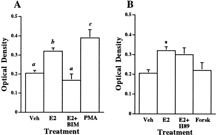
The oxytocin receptor (OTR) is differentially expressed in the CNS. Because there are multiple mechanisms by which the OTR can be transcriptionally induced, we hypothesized that differences in OTR expression may be explained by activation of distinct signal transduction pathways and may be critical for the control of anxiety and sex behaviors. To determine the regulation and functional significance of this expression, we infused female rats with modifiers of protein kinases before assaying for behavior and oxytocin receptor binding. In the ventromedial nucleus of the hypothalamus (VMH), estrogen-dependent induction of oxytocin receptors required protein kinase C activation, and oxytocin infused here promoted female sex behavior but had no effect on anxiety. In contrast, dopamine controlled tonic oxytocin receptor expression in the central nucleus of the amygdala (cAmyg) through activation of protein kinase A, and oxytocin infused here was anxiolytic but had no effect on female sex behavior. Therefore, we have identified brain region-specific regulation of the OTR in the VMH and cAmyg. Distinct signal transduction pathways regulating receptor expression and binding in each brain region may mediate in part the ability of oxytocin to exert these differential behavioral effects.


Figures

Fig. 1.
Fig. 1.
Representative autoradiographs illustrating oxytocin receptor binding in the VMH. Animals were pretreated with estradiol (2 μg) or oil vehicle by subcutaneous injection; infused centrally, directly into the VMH with BIM (inhibitor of PKC) or the phorbol ester, PMA, 8 hr before collection of the brains; and assayed for OTR binding by quantitative receptor autoradiography. Treatment groups were as follows: A, subcutaneous oil vehicle injection, saline vehicle infusion into the VMH;B, subcutaneous estradiol injection, saline vehicle infusion into the VMH; C, subcutaneous estradiol injection, BIM infusion into the VMH; D, subcutaneous oil vehicle injection, PMA infusion into the VMH. Arrowindicates region of VMH that was analyzed.
Fig. 2.
Fig. 2.
Quantification of OTR binding in the VMH.A, Optical density levels of OTR binding in the VMH of ovariectomized rats treated peripherally with estradiol (E2) or oil vehicle and centrally with inhibitors (BIM) or activators (PMA) of PKC. Treatment groups were Veh (subcutaneous oil vehicle injection, saline vehicle infusion into the VMH; n= 4), E2 [subcutaneous estradiol (2 μg) injection, saline vehicle infusion into the VMH; n = 7],E2 + BIM [subcutaneous estradiol injection, BIM (200 μm) infusion into the VMH; n = 4], or PMA [subcutaneous oil vehicle injection, PMA (200 nm) infusion into the VMH; n = 5]. Brains were collected 8 hr after drug infusion and steroid treatment. Data were analyzed by one-way ANOVA and indicate a significant effect of treatment [F(3,16) = 12.01;p < 0.001]; post hoc analysis indicates that a is significantly different fromb (p < 0.01) andc (p < 0.001).B, When compared with the same Veh- and E2-treated groups as above, infusion of forskolin (Forsk) (200 nm; n = 3), an activator of PKA, or H89 (E2 + H89) (200 μm;n = 3), an inhibitor of PKA, into the VMH of ovariectomized females was without effect on OTR binding in the VMH. All values are ± SEM.
Fig. 3.
Fig. 3.
Representative autoradiographs illustrating OTR binding in the cAmyg. Animals were infused directly into the cAmyg, and brains were collected 8 hr later and assayed for OTR binding by quantitative receptor autoradiography. Treatments were as follows:A, saline vehicle infusion into the cAmyg;B, H89 (PKA inhibitor) infusion into the cAmyg;C, SCH23390 (dopamine D1 receptor antagonist) infusion into the cAmyg; or D, SCH23390 + forskolin infusion into the cAmyg. Arrow indicates region of cAmyg that was analyzed.
Fig. 4.
Fig. 4.
Quantification of OTR binding in the cAmyg. Ovariectomized rats were treated as follows: A,Veh 1 (saline vehicle for H89 infusion into the cAmyg;n = 7), and H89 (H89 infusion into cAmyg; n = 5); B, Veh2 (saline vehicle for SCH23390 infusion into the cAmyg;n = 2); SCH (SCH23390 infusion into the cAmyg; n = 3), or SCH + Forsk(SCH23390 + forskolin infusion into the cAmyg; n = 3). Brains were collected 8 hr after drug infusion. There was a significant effect of H89 infusion [ANOVA;F(1,10) = 13.96] and SCH23390 infusion [F(3,7) = 10.62; p< 0.01] when each was compared with its own controls. Post hoc analysis, **p < 0.01. All values are ± SEM. Previous treatment with estradiol was found to have no influence on the effectiveness of H89 treatment (data not shown).
Fig. 5.
Fig. 5.
Effects of oxytocin infusion into the VMH and cAmyg on female sex behavior. Animals were primed with low doses of estradiol for 2 d and then pretested for lordosis responding before infusion of oxytocin (OT; 1 μg) or saline (Sal) into the VMH or cAmyg. Post-testing was performed 10–20 min after infusion. Data are presented as the increase in lordosis amplitude between the pretest and post-test.A, For animals with indwelling cannulas in the VMH, oxytocin infusion significantly increased lordosis responding compared with animals either infused with saline or pretreated for 2 d with the PKC inhibitor, BIM. ANOVA with post hoc Kruskal—Wallis; **p < 0.001;n = 9 for all groups. These results show that oxytocin infusion into the VMH increases lordosis behavior and this behavior is blocked by pretreatment with BIM. B, For animals with indwelling cannulas in the cAmyg, there was no difference in lordosis responding between those infused with oxytocin (n = 5) versus saline [n = 4;F(1,14) = 0.25; p> 0.5]. All values are ± SEM.
Fig. 6.
Fig. 6.
Effects of oxytocin infusion into the VMH and cAmyg on activity in an open field. Animals were placed into the center of an open-field testing apparatus within 10–20 min of infusion of oxytocin (1 μg) or saline. The ratio of inside/outside crossings is a useful overall indicator of activity in an open field. An increase in the ratio indicates reduced anxiety-like behavior because it demonstrates that the animals are more willing to explore the center of the open field. A, For animals with indwelling cannulas in the cAmyg and infused with oxytocin (n = 9), there was a significant increase in the ratio when compared with saline-infused controls (n = 6). This effect was blocked by pretreatment with the dopamine D1 receptor antagonist, SCH23390 (n = 8) (**p < 0.01; ANOVA). There were no differences in overall activity between any groups. B, For animals with indwelling cannulas in the VMH, there was no effect of oxytocin infusion on activity in the open field.
Fig. 7.
Fig. 7.
Summary diagram of effects observed on oxytocin receptor binding by estradiol and modulators of PKC and PKA and the resulting effects on behavior. Estradiol increases oxytocin receptor binding selectively in the VMN but not in the cAmyg. Estrogen action in the VMN involves PKC and results in increased female sex behavior as demonstrated by increased lordosis. Dopamine activation of the D1 receptor increases activation of PKA, resulting in increased or tonically maintained levels of oxytocin receptor binding in the cAmyg. Oxytocin acting in the cAmyg decreases anxiety. The use of distinct signal transduction pathways to regulate oxytocin receptor levels in the VMN and cAmyg may have evolved to allow for constitutive expression of oxytocin receptors in the cAmyg and an immediate response to possible life-threatening stimuli, as opposed to the hormonally constrained control of oxytocin receptors in the VMN that regulate female reproductive behavior.

References

    1. Arletti R, Bertolini A. Oxytocin acts as an antidepressant in two animal models of depression. Life Sci. 1987;41:1725–1730. - PubMed
    1. Bale TL, Dorsa DM. Sex differences in and effects of estrogen on oxytocin receptor messenger ribonucleic acid expression in the ventromedial hypothalamus. Endocrinology. 1995a;136:27–32. - PubMed
    1. Bale TL, Dorsa DM. Regulation of oxytocin receptor messenger ribonucleic acid in the ventromedial hypothalamus by testosterone and its metabolites. Endocrinology. 1995b;136:5135–5138. - PubMed
    1. Bale TL, Dorsa DM. Cloning, novel promoter sequence, and estrogen regulation of a rat oxytocin receptor gene. Endocrinology. 1997;138:1151–1158. - PubMed
    1. Bale TL, Dorsa DM. NGF, cyclic AMP, and phorbol esters regulate oxytocin receptor gene transcription in SK-N-SH and MCF7 cells. Mol Brain Res. 1998;53:130–137. - PubMed

Publication types

MeSH terms