. 2001 Apr;75(8):3520-6.
doi: 10.1128/JVI.75.8.3520-3526.2001.

Two functionally distinct forms of a retroviral receptor explain the nonreciprocal receptor interference among subgroups B, D, and E avian leukosis viruses

Affiliations

Two functionally distinct forms of a retroviral receptor explain the nonreciprocal receptor interference among subgroups B, D, and E avian leukosis viruses

H B Adkins et al. J Virol. 2001 Apr.

Abstract

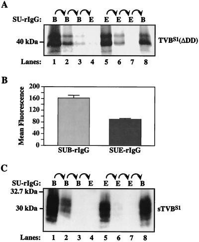
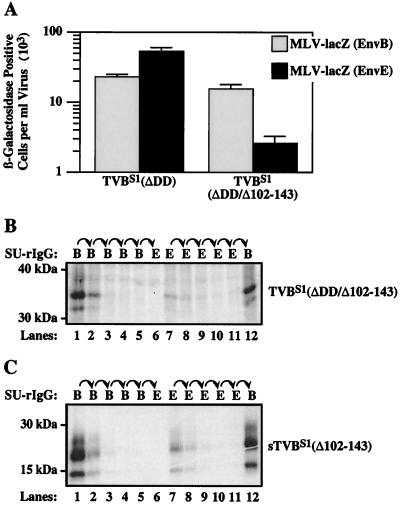
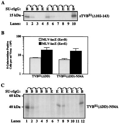
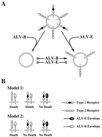
Subgroups B, D, and E avian leukosis viruses (ALV-B, -D, and -E) share the same chicken receptor, TVB(S1), a tumor necrosis factor receptor (TNFR)-related protein. These viruses, however, exhibit nonreciprocal receptor interference (NRI): cells preinfected with ALV-B or ALV-D are resistant to superinfection by viruses of all three subgroups, whereas those pre-infected by ALV-E are resistant only to superinfection by other subgroup E viruses. In this study, we investigated the basis of this phenomenon by characterizing the interaction of TVB(S1) with ALV-B Env or ALV-E Env. Sequential immunoprecipitation analysis using surface envelope immunoglobulin fusion proteins revealed the existence of two separate types of TVB(S1) that are encoded by the same cDNA clone. One form, designated the type 1 receptor, is specific for ALV-B and ALV-E. The other form, the type 2 receptor, is specific for ALV-B. We show that a protein consisting of only the first and second extracellular cysteine-rich domains of TVB(S1) is capable of forming both receptor types. However, the third extracellular cysteine-rich domain is required for efficient formation of the type 1 receptor. We also demonstrate that heterogeneous N-linked glycosylation cannot explain the difference in activities of the two receptor types. The existence of two types of TVB(S1) explains the NRI pattern between ALV-B and -E: subgroup B viruses establish receptor interference with both receptor types, whereas subgroup E viruses interfere only with the type 1 receptor, leaving the type 2 receptor available to mediate subsequent rounds of ALV-B entry. The formation of a TVB receptor type that is specific for cytopathic ALV may also have important implications for understanding how some subgroups of ALV cause cell death.


Figures

FIG. 1
FIG. 1
Schematic diagram of the TVBS1 constructs used in these studies. The TVBS1 and TVBS1 (ΔDD) proteins were described previously (2). The other TVB proteins were generated specifically for these experiments. The amino acid residues are numbered according to a scheme used previously (6). SP, signal peptide; TM, transmembrane region; DD, cytoplasmic death domain.
FIG. 2
FIG. 2
TVBS1 is produced as two distinct receptor types. (A) Aliquots of [35S]cysteine-labeled protein lysate prepared from transfected human 293 cells that express TVBS1(ΔDD) were subjected to three rounds of sequential immunoprecipitation with protein A-Sepharose beads loaded with either SUB-rIgG (lanes 1 to 3) or SUE-rIgG (lanes 5 to 7) and then with SUE-rIgG (lane 4) or SUB-rIgG (lane 8). The precipitated TVB proteins were visualized by autoradiography. (B) Flow cytometric analysis of 293 cells expressing TVBS1 (ΔDD) was performed with SUB-rIgG or SUE-rIgG, followed by a fluorescein isothiocyanate-conjugated secondary antibody. The results of a representative experiment performed in triplicate are shown as the mean fluorescence values of each cell population with the standard deviations of the data indicated with error bars. (C) An experiment similar to that described for panel A was performed with extracellular supernatant containing sTVBS1. In this case, the immunoprecipitated proteins were detected by immunoblotting with SUB-rIgG and an HRP-conjugated secondary antibody followed by enhanced chemiluminescence.
FIG. 3
FIG. 3
The first two CRDs of TVBS1 are sufficient for formation of the type 1 receptor. (A) Human 293 cells were transiently transfected with a plasmid expressing TVBS1(ΔDD) or TVBS1(ΔDD/Δ102-143) and then challenged with MLV-LacZ (Env B) or MLV-LacZ (Env E). Infected cells were identified by staining for β-galactosidase activity. The data from three independent experiments with standard deviations are shown. Aliquots of [35S]cysteine-labeled protein lysate containing TVBS1(ΔDD/Δ102-143) (B) or supernatant containing sTVBS1 (Δ102-143) (C) were subjected to the sequential immunoprecipitation procedure outlined for Fig. 2A, and the precipitated proteins were visualized by autoradiography following SDS-polyacrylamide gel electrophoresis.
FIG. 4
FIG. 4
The existence of both receptor types cannot be explained by heterogeneous N-linked glycosylation. (A) An aliquot of [35S]cysteine labeled extracellular supernatant containing sTVBS1(Δ102-143) was incubated with N-glycosidase F and neuraminidase and then subjected to serial rounds of immunoprecipitation with SUB-rIgG and SUE-rIgG. The precipitated proteins were subjected to SDS-polyacrylamide gel electrophoresis and visualized by autoradiography. (B) Human 293 cells expressing TVBS1 (ΔDD) and TVBS1(ΔDD)-N54A were challenged with MLV-LacZ (Env B) or MLV-LacZ (Env E) and infected cells were identified and counted after staining for β-galactosidase activity. The data shown represents the average values obtained in three independent experiments with the standard deviations given by error bars. (C) An aliquot of [35S]cysteine-labeled protein lysate containing TVBS1(ΔDD)-N54A was subjected to the sequential immunoprecipitation assay, and proteins were visualized by autoradiography following SDS-polyacrylamide gel electrophoresis.
FIG. 5
FIG. 5
Implications of the two receptor types for understanding NRI between ALV-B and ALV-E and for ALV-induced cytopathic effects. (A) NRI between ALV-B and ALV-E; (B) possible role of TVBS1 in cell death induced by ALV-B. These models are discussed in detail in the text.

Similar articles

Cited by

References

    1. Adkins H B, Brojatsch J, Naughton J, Rolls M M, Pesola J M, Young J A T. Identification of a cellular receptor for subgroup E avian leukosis virus. Proc Natl Acad Sci USA. 1997;94:11617–11622. - PMC - PubMed
    1. Adkins H B, Brojatsch J, Young J A T. Identification and characterization of a shared TNFR-related receptor for subgroup B, D, and E avian leukosis viruses reveal cysteine residues required specifically for subgroup E viral entry. J Virol. 2000;74:3572–3578. - PMC - PubMed
    1. Battini J L, Rasko J E, Miller A D. A human cell-surface receptor for xenotropic and polytropic murine leukemia viruses: possible role in G protein-coupled signal transduction. Proc Natl Acad Sci USA. 1999;96:1385–1390. - PMC - PubMed
    1. Boeke J D, Stoye J P. Retrotransposons, endogenous retroviruses, and the evolution of retroelements. In: Coffin J M, Hughes S H, Varmus H E, editors. Retroviruses. Cold Spring Harbor, N.Y: Cold Spring Harbor Laboratory Press; 1997. pp. 343–435. - PubMed
    1. Brojatsch J, Naughton J, Adkins H B, Young J A T. TVB receptors for cytopathic and noncytopathic subgroups of avian leukosis viruses are functional death receptors. J Virol. 2000;74:11490–11494. - PMC - PubMed

Publication types

MeSH terms

LinkOut - more resources