. 2001 Apr;75(8):3527-36.
doi: 10.1128/JVI.75.8.3527-3536.2001.

Antiviral effect of N-butyldeoxynojirimycin against bovine viral diarrhea virus correlates with misfolding of E2 envelope proteins and impairment of their association into E1-E2 heterodimers

Affiliations

Antiviral effect of N-butyldeoxynojirimycin against bovine viral diarrhea virus correlates with misfolding of E2 envelope proteins and impairment of their association into E1-E2 heterodimers

N Branza-Nichita et al. J Virol. 2001 Apr.

Abstract

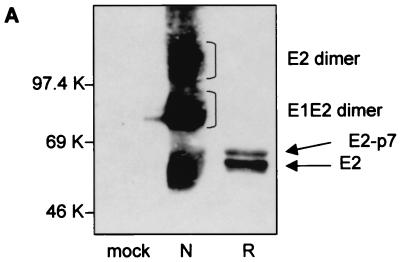
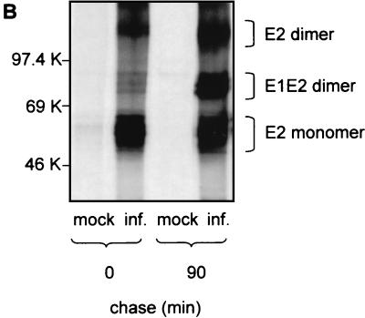
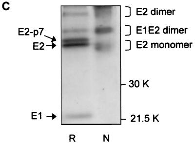
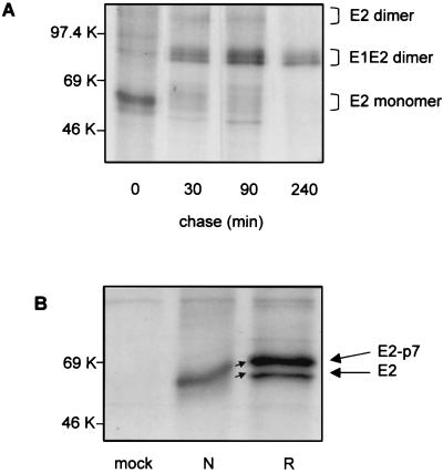
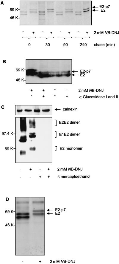
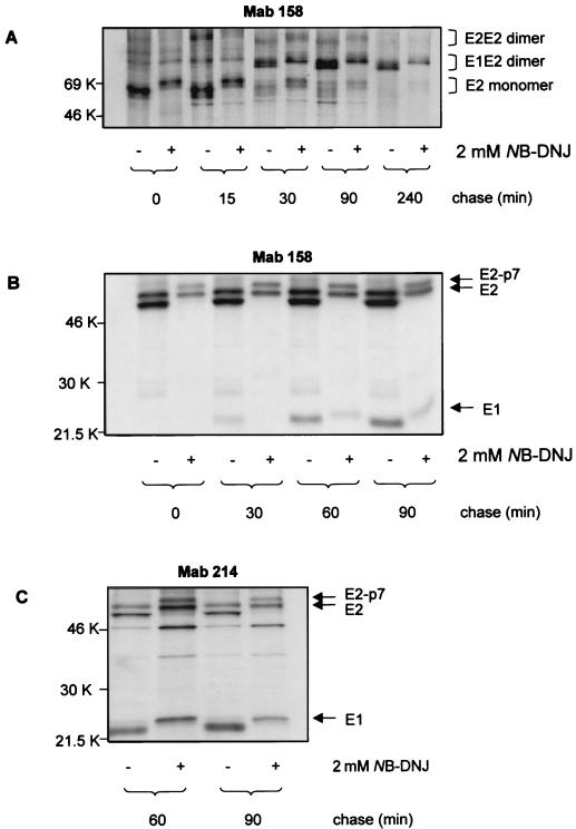
The iminosugar N-butyldeoxynojirimycin (NB-DNJ), an endoplasmic reticulum alpha-glucosidase inhibitor, has an antiviral effect against bovine viral diarrhea virus (BVDV). In this report, we investigate the molecular mechanism of this inhibition by studying the folding pathway of BVDV envelope glycoproteins in the presence and absence of NB-DNJ. Our results show that, while the disulfide-dependent folding of E2 glycoprotein occurs rapidly (2.5 min), the folding of E1 occurs slowly (30 min). Both BVDV envelope glycoproteins associate rapidly with calnexin and dissociate with different kinetics. The release of E1 from the interaction with calnexin coincides with the beginning of E1 and E2 association into disulfide-linked heterodimers. In the presence of NB-DNJ, the interaction of E1 and E2 with calnexin is prevented, leading to misfolding of the envelope glycoproteins and inefficient formation of E1-E2 heterodimers. The degree of misfolding and the lack of association of E1 and E2 into disulfide-linked complexes in the presence of NB-DNJ correlate with the dose-dependent antiviral effect observed for this iminosugar.


Figures

FIG. 1
FIG. 1
Analysis of BVDV envelope glycoproteins expressed in MBDK cells. (A) MBDK cells were infected with BVDV (NADL strain) at an MOI of 1. Eighteen hours p.i. the cells were lysed and analyzed by SDS–10% PAGE under nonreducing (lane N) and reducing (lane R) conditions, followed by Western blot analysis with MAb 214. (B) MBDK cells were infected with BVDV at an MOI of 1. Eighteen hours p.i. the cells were pulse-labeled with [35S]methionine and [35S]cysteine for 15 min, chased for the times indicated, lysed, and immunoprecipitated with MAb 214. Immunoprecipitated proteins were separated by SDS–10% PAGE under nonreducing conditions and analyzed by autoradiography. (C) Conditions were the same as described for panel B, except that the sample at min 90 of chase was analyzed by SDS–12% PAGE, under both nonreducing and reducing conditions. Mock-infected cells (mock inf.) were included as controls.
FIG. 1
FIG. 1
Analysis of BVDV envelope glycoproteins expressed in MBDK cells. (A) MBDK cells were infected with BVDV (NADL strain) at an MOI of 1. Eighteen hours p.i. the cells were lysed and analyzed by SDS–10% PAGE under nonreducing (lane N) and reducing (lane R) conditions, followed by Western blot analysis with MAb 214. (B) MBDK cells were infected with BVDV at an MOI of 1. Eighteen hours p.i. the cells were pulse-labeled with [35S]methionine and [35S]cysteine for 15 min, chased for the times indicated, lysed, and immunoprecipitated with MAb 214. Immunoprecipitated proteins were separated by SDS–10% PAGE under nonreducing conditions and analyzed by autoradiography. (C) Conditions were the same as described for panel B, except that the sample at min 90 of chase was analyzed by SDS–12% PAGE, under both nonreducing and reducing conditions. Mock-infected cells (mock inf.) were included as controls.
FIG. 2
FIG. 2
Analysis of intra- and intermolecular disulfide bond formation of BVDV envelope glycoproteins. MBDK cells were infected with BVDV at an MOI of 1. Eighteen hours p.i., the cells were either pulse-labeled with [35S]methionine and [35S]cysteine for 15 min and chased for the times indicated (A) or pulse-labeled for 2.5 min and harvested immediately (B). Cell lysates were immunoprecipitated with MAb 214, and proteins bound were analyzed by SDS–10% PAGE under nonreducing conditions (A) and both nonreducing (lane N) and reducing (lane R) conditions (B). Mock-infected cells (mock) were included as controls.
FIG. 3
FIG. 3
Interaction of E1 and E2 BVDV glycoproteins with calnexin. MDBK cells were either infected with BVDV at an MOI of 1 (inf.) or mock infected (mock). (A and C) Eighteen hours p.i., cells were pulse-labeled for 2.5 min with [35S]methionine and [35S]cysteine, chased for the times indicated, and either immunoprecipitated with MAb 214 and polyclonal anticalnexin antibodies (α cal) alone or coimmunoprecipitated with both MAb 214 and α cal, as indicated. The immunoprecipitated proteins were analyzed by SDS–10% (A) or SDS–12% PAGE under reducing conditions and visualized by autoradiography (C, graph). Calnexin-bound E1 was quantified by densitometric analysis (C, graph), with the amount of E1 associated with calnexin at time point 0 equaling 100%. (B) Fractions of the cell lysates were analyzed either before immunoprecipitation (lanes 3 and 4) or following immunoprecipitation with MAb 214 and elution of bound proteins under reducing conditions. The proteins were separated by SDS–10% PAGE and analyzed with a Western blot stained with α cal.
FIG. 4
FIG. 4
Biosynthesis and processing of BVDV envelope glycoproteins in the presence of NB-DNJ. MDBK cells were infected with BVDV at an MOI of 1. (A) Eighteen hours p.i., the cells were treated (+) or not treated (−) with 2 mM NB-DNJ. Two hours later, the cells were pulse-labeled with [35S]methionine and [35S]cysteine for 15 min, chased for the times indicated in the continuous presence of the drug, and immunoprecipitated with MAb 214. The proteins were analyzed by SDS–10% PAGE under reducing conditions and visualized by autoradiography. (B) Infected cells were grown for 18 h in the absence (−) or presence (+) of 2 mM NB-DNJ. Cell lysates were analyzed for protein content, and the equivalent of 20 μg of protein was digested (+) or not digested (−) with a mixture of α-glucosidases I and II. The proteins were separated by SDS–10% PAGE under reducing conditions and analyzed with a Western blot stained with MAb 214. (C) Infected cells were grown for 18 h in the absence (−) or presence (+) of 2 mM NB-DNJ. Cell lysates were boiled for 5 min in the absence (−) or presence (+) of 5% β-mercaptoethanol prior to separation by SDS–10% PAGE. The proteins were analyzed with a Western blot stained with MAb 158. Calnexin was detected with an anticalnexin antibody and used as loading control. (D) Conditions were the same as described for panel A, except that infected cells were lysed immediately after the 15 min of pulse and immunoprecipitated with MAb 158.
FIG. 5
FIG. 5
Folding and assembly of E1 and E2 glycoproteins in NB-DNJ-treated cells. (A and B) MDBK cells were infected with BVDV at an MOI of 1. Eighteen hours p.i., the cells were treated (+) or not treated (−) with 2 mM NB-DNJ for 2 h before being pulse-labeled with [35S]methionine and [35S]cysteine for 15 min. At the chase times indicated, the cells were lysed and immunoprecipitated with MAb 158. Proteins bound were separated by SDS–10% PAGE under nonreducing conditions (A) or by SDS–12% PAGE under reducing conditions (B) and visualized by autoradiography. (C) Conditions were the same as those described for panel B, except that the cell lysates were immunoprecipitated with MAb 214.
FIG. 6
FIG. 6
BVDV protein folding and infectivity in NB-DNJ-treated cells. (A) MDBK cells were grown to subconfluence in six-well plates and infected with BVDV at an MOI of 1. After 1 h the inoculum was removed and the cells were grown for 24 h in medium containing either NB-DNJ or NB-DGJ at the indicated concentrations (plaque assay). The supernatants containing secreted virus were then used to infect fresh MDBK monolayers, and the plaques were counted after 24 h (yield assay). Results are the percentages of the number of plaques resulting from infection with the inhibitor-free plaque assay supernatant (considered as 100%). (B) MDBK cells were infected with BVDV at an MOI of 1 and grown in the absence or presence of increasing concentrations of NB-DNJ. Eighteen hours p.i., the cells were labeled for 15 min with [35S]methionine and [35S]cysteine and lysed. The amount of radiolabeled protein in the cell lysates was adjusted to 3 × 10⁶ cpm/ml, before immunoprecipitation with MAb 158 and antiactin antibodies (loading control). The amount of immunoprecipitated protein was quantified by liquid scintillation counting and expressed as the percentage of counts per minute determined for the drug-free samples. (C) MDBK cells were infected with BVDV at an MOI of 1 and grown in the absence or presence of NB-DNJ at the concentrations indicated. Eighteen hours p.i., the cells were lysed and the assembly of the viral proteins was analyzed by SDS–10% PAGE under nonreducing conditions followed by Western blotting using MAb 214. Calnexin and actin were detected on the same membrane (loading control) (gels). The band intensities of E2 monomers as well as of E1-E2 and E2-E2 dimers were quantified by densitometric analysis (graph).

References

    1. Baginski S G, Peaver D C, Seipel M, Sun S C C, Benetatos C A, Chunduru S K, Rice C M, Collet M S. Mechanism of action of a pestivirus antiviral compound. Proc Natl Acad Sci USA. 2000;97:7981–7986. - PMC - PubMed
    1. Bergeron J J M, Brenner M B, Thomas D Y, Williams D B. Calnexin: a membrane-bound chaperone of the endoplasmic reticulum. Trends Biochem Sci. 1994;19:124–129. - PubMed
    1. Block T M, Lu X, Platt F M, Foster G R, Gerlich W H, Blumberg B S, Dwek R A. Secretion of human hepatitis B virus is inhibited by the imino sugar N-butyldeoxynojirimycin. Proc Natl Acad Sci USA. 1994;91:2235–2239. - PMC - PubMed
    1. Braakman I, Helenius J, Helenius A. Manipulating disulfide bond formation and protein folding in the endoplasmic reticulum. EMBO J. 1992;11:1717–1722. - PMC - PubMed
    1. Choukhi A, Ung S, Wychowsky C, Dubuisson J. Involvement of endoplasmic reticulum chaperones in the folding of hepatitis C virus glycoproteins. J Virol. 1998;72:3851–3858. - PMC - PubMed

Publication types

MeSH terms

LinkOut - more resources