. 2001 Apr;75(8):3626-35.
doi: 10.1128/JVI.75.8.3626-3635.2001.

Characterization of intracellular reverse transcription complexes of human immunodeficiency virus type 1

Affiliations

Characterization of intracellular reverse transcription complexes of human immunodeficiency virus type 1

A Fassati et al. J Virol. 2001 Apr.

Abstract

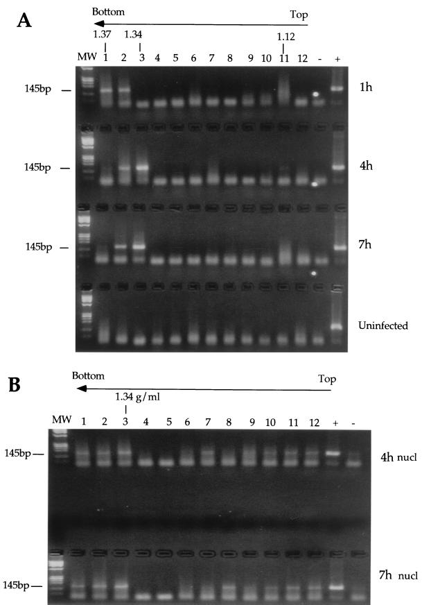
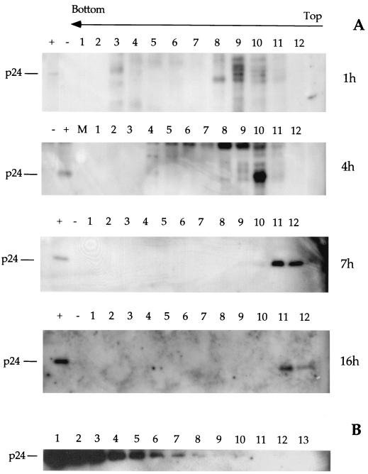
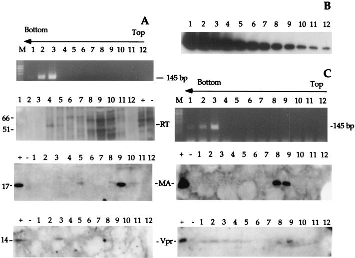
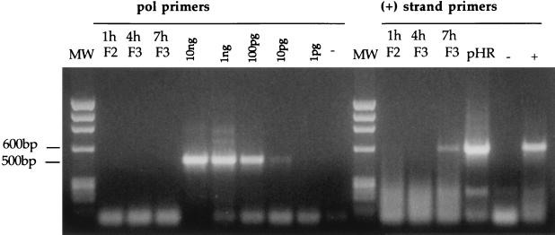
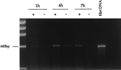
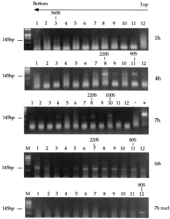
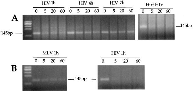
To examine the early events of the life cycle of human immunodeficiency virus type 1 (HIV-1), we analyzed the intracellular complexes mediating reverse transcription isolated from acutely infected cells. Partial purification of the reverse transcription complexes (RTCs) by equilibrium density fractionation and velocity sedimentation indicated that two species of RTCs are formed but only one species is able to synthesize DNA. Most of the capsid, matrix, and reverse transcriptase (RT) proteins dissociate from the complex soon after cell infection, but Vpr remains associated with the RTC. The RTCs isolated 1, 4, and 7 h after infection are competent for reverse transcription in vitro, indicating that a small proportion of RT remains associated with them. HIV RTCs isolated early after infection have a sedimentation velocity of approximately 560S. Later, different species with a sedimentation velocity ranging from 350S to 100S appear. Nuclear-associated RTCs have a sedimentation velocity of 80S. Shortly after initiation of reverse transcription, the viral strong-stop DNA within the RTC is sensitive to nuclease digestion and becomes protected when reverse transcription is almost completed.


Figures

FIG. 1
FIG. 1
PCR analyses of cytoplasmic (A) and nuclear (B) extracts after equilibrium density fractionation using primers specific for the strong-stop DNA (expected band size is 145 bp). HeLa cells were infected with an HIV-1-based vector; cell extracts were prepared 1, 4, and 7 h postinfection, loaded on a 20 to 70% linear sucrose gradient, and centrifuged at 4°C for 20 h at 35,000 rpm in a Beckman SW55 rotor. After centrifugation, gradients were collected in 12 fractions and analyzed. Arrows indicate the direction of the gradient from the lowest (top) to the highest (bottom) density. The density of the fraction containing the peak of the viral DNA is indicated for each time point. Lanes: MW, DNA molecular weight standards; 1 to 12, fractions 1 to 12. The rapidly migrating bands are PCR artifacts. Hirt viral DNA was used as positive control (+); uninfected Hela cells served as a negative control (−). nucl, nuclear.
FIG. 2
FIG. 2
(A) Western blot analyses of equilibrium density fractions containing cytoplasmic extracts collected 1, 4, 7, and 16 h postinfection. Following SDS-PAGE and protein transfer, the PVDF membrane was probed with a monoclonal antibody against HIV p24 capsid protein. Lanes: 1 to 12, fractions 1 to 12 as in Fig. 1; +, purified virus; −, uninfected HeLa cells; M, molecular weight markers. The arrow indicates the direction of the gradient from the lowest (top) to the highest (bottom) density. (B) Testing of sensitivity of Western blotting using recombinant p24. Following SDS-PAGE and protein transfer, the PVDF membrane was probed with the same antibody as used for panel A and for the same exposure time. Lanes 1 to 13 correspond to twofold serial dilutions of recombinant p24 from 3.2 ng to 0.7 pg.
FIG. 3
FIG. 3
(A and C) PCR and Western blot analyses of equilibrium density fractions containing cytoplasmic extracts collected 4 (A) and 16 (C) h after acute infection. Fractions were subjected to PCR with primers specific for the strong-stop DNA (expected size is 145 bp). Lanes: M, DNA molecular weight standards; 1 to 12, fractions 1 to 12. Following SDS-PAGE and protein transfer, the PVDF membrane was probed with a polyclonal antibody against Vpr and a monoclonal antibody against MA and RT. Controls were purified HIV-1 (+) and conditioned medium from untransfected 293T cells (−). Arrows indicate the direction of the gradient, from the lowest density (top) to the highest density (bottom). Positions of molecular weight markers are indicated in kilodaltons on the left. (B) Testing of sensitivity of Western blotting using recombinant p17. Following SDS-PAGE and protein transfer, the PVDF membrane was probed with the same antibody as used for panel A and for the same exposure time. Lanes 1 to 12 correspond to twofold serial dilutions of recombinant p17 from 8 ng to 3.5 pg.
FIG. 4
FIG. 4
PCR analyses of equilibrium density fractions containing cytoplasmic extracts collected 1, 4, and 7 h postinfection. Selected fractions from the equilibrium density gradients shown in Fig. 1 were subjected to PCR using primers specific for the Gag-Pol region (pol primers; expected band size is 500 bp) and for the positive (+)-strand DNA (expected size is 600 bp). Fractions 2 and 3 (F2 and F3) are the same as in Fig. 1. Serial dilutions of pCMVΔR9 plasmid DNA were used as the internal standard for Pol PCR. Hirt DNA from uninfected cells was used as negative control (−). Plasmid pHR' and Hirt DNA from infected cells were used as internal controls for the (+)-strand PCR. MW, DNA molecular weight standards. Low-molecular-weight bands are PCR artifacts.
FIG. 5
FIG. 5
Endogenous reverse transcription assay of equilibrium density fractions containing the peak of the viral DNA from cytoplasmic extracts collected 1, 4, and 7 h postinfection. Samples diluted 1:10 were incubated for 6 h at 37°C in the presence (+) or absence (−) exogenous dNTPs and then subjected to PCR with primers specific for the (+)-strand DNA (expected band size is 600 bp).
FIG. 6
FIG. 6
Analysis of RTCs by sedimentation velocity. Cytoplasmic extracts collected 1, 4, 7, and 16 h postinfection and nuclear extracts collected 7 h postinfection were subjected to equilibrium density centrifugation. The fractions containing the peak of the retroviral DNA were further purified, concentrated using a Centricon filter, and centrifuged through a 5 to 20% linear sucrose gradient for 1 h at 23,000 rpm in a Beckman SW55 rotor. Twelve fractions were collected, and the viral DNA in each fraction was detected by PCR using primers specific for the strong-stop DNA (expected band size is 145 bp). The rapidly migrating bands are PCR artifacts. The arrow indicates the direction of the gradient. nucl, nuclear.
FIG. 7
FIG. 7
Sensitivity of the viral strong-stop DNA within the RTC to micrococcal nuclease digestion. (A) Equilibrium density fractions containing the peak of the viral DNA from the cytoplasmic extracts collected 1, 4, and 7 h postinfection were incubated on ice in the presence of micrococcal nuclease and 2 mM CaCl2. The reactions were stopped by addition of 4 mM EGTA at the indicated time points and analyzed by PCR using primers specific for the strong-stop DNA (expected band size is 145 bp). Naked viral DNA was incubated as above in the presence of micrococcal nuclease and cytoplasmic extracts from uninfected cells (Hirt HIV). (B) Equilibrium density fractions containing the peak of HIV-1 and MoMLV DNAs were mixed 1:1 and subjected to micrococcal nuclease digestion as above. The reaction was analyzed by PCR using primers specific for HIV or MoMLV strong stop. To ensure the linearity of amplification, the samples were subjected to two independent amplification rounds, one of 30 and one of 40 cycles.

Similar articles

Cited by

References

    1. Bukrinskaya A, Brichacek B, Mann A, Stevenson M. Establishment of a functional human immunodeficiency virus type 1 (HIV-1) reverse transcription complex involves the cytoskeleton. J Exp Med. 1998;188:2113–2125. - PMC - PubMed
    1. Bukrinsky M I, Sharova N, Dempsey M P, Stanwick T L, Bukrinkaya A G, Haggerty S, Stevenson M. Active nuclear import of human immunodeficiency virus type 1 preintegration complexes. Proc Natl Acad Sci USA. 1992;89:6580–6584. - PMC - PubMed
    1. Bukrinsky M I, Haggerty S, Dempsey M I, Sharova N, Adzhubel A, Spitz L, Lewis P, Goldfarb D, Emerman M, Stevenson M. A nuclear localization signal within HIV-1 matrix protein that governs infection of non-dividing cells. Nature. 1993;365:666–669. - PMC - PubMed
    1. Bukrinsky M I, Sharova N, McDonald T L, Pushkarskaya T, Tarpley W G, Stevenson M. Association of integrase, matrix and reverse transcriptase antigens of human immunodeficiency virus type 1 with viral nucleic acids following acute infection. Proc Natl Acad Sci USA. 1993;90:6125–6129. - PMC - PubMed
    1. Farnet C M, Bushman F D. HIV-1 cDNA integration: requirement of HMG I(Y) protein for function of preintegration complexes in vitro. Cell. 1997;88:483–492. - PubMed

Publication types

MeSH terms

LinkOut - more resources