. 2001 Apr;75(8):3753-65.
doi: 10.1128/JVI.75.8.3753-3765.2001.

Route of simian immunodeficiency virus inoculation determines the complexity but not the identity of viral variant populations that infect rhesus macaques

Affiliations

Route of simian immunodeficiency virus inoculation determines the complexity but not the identity of viral variant populations that infect rhesus macaques

J L Greenier et al. J Virol. 2001 Apr.

Abstract

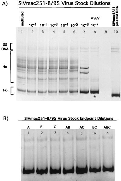
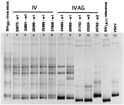
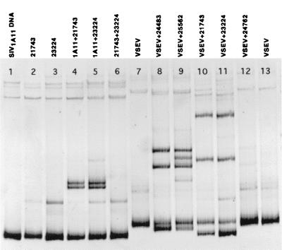
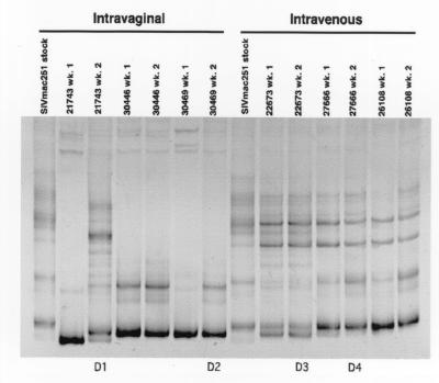
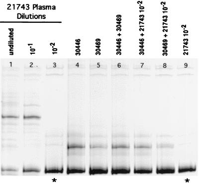
A better understanding of the host and viral factors associated with human immunodeficiency virus (HIV) transmission is essential to developing effective strategies to curb the global HIV epidemic. Here we used the rhesus macaque-simian immunodeficiency virus (SIV) animal model of HIV infection to study the range of viral genotypes that are transmitted by different routes of inoculation and by different types of viral inocula. Analysis of transmitted variants was undertaken in outbred rhesus macaques inoculated intravenously (IV) or intravaginally (IVAG) with a genetically heterogeneous SIVmac251 stock derived from a well-characterized rhesus macaque viral isolate. In addition, we performed serial IV and IVAG passage experiments using plasma from SIV-infected macaques as the inoculum. We analyzed the V1-V2 region of the SIV envelope gene from virion-associated RNA in plasma from infected animals by the heteroduplex mobility assay (HMA) and by DNA sequence analysis. We found that a more diverse population of SIV genetic variants was present in the earliest virus-positive plasma samples from all five IV SIVmac251-inoculated monkeys and from two of five IVAG SIVmac251-inoculated monkeys. In contrast, we found a relatively homogeneous population of SIV envelope variants in three of five monkeys inoculated IVAG with SIVmac251 stock and in two monkeys infected after IVAG inoculation with plasma from an SIV-infected animal. In some IVAG-inoculated animals, the transmitted SIV variant was the most common variant in the inoculum. However, a specific viral variant in the SIVmac251 stock was not consistently transmitted by IVAG inoculation. Thus, it is likely that host factors or stochastic processes determine the specific viral variants that infect an animal after IVAG SIV exposure. In addition, our results clearly demonstrate that the route of inoculation is associated with the extent and breadth of the genetic complexity of the viral variant population in the earliest stages of systemic infection.


Figures

FIG. 1
FIG. 1
SIV V1-V2 variants present in serial dilutions of SIVmac251-8/95 virus stock. (A) Lanes 1 to 8, dilutions of virus stock from undiluted to 10−7. Heteroduplex (He) bands indicate the presence of multiple V1-V2 variants in the virus stock. The last dilution yielding a RT-PCR product (10−7) was comprised of a homogeneous variant population (i.e., a single variant) as depicted by the presence of a single homoduplex (Ho) band in lane 8. The V1-V2 sequence amplified from the 10−7 dilution (∗) was considered to be the most common envelope variant in the virus stock; this variant was designated VSEV. Lane 9, no sample loaded. Lane 10 contains the V1-V2 fragment of SIV amplified from SIVmac1A11 plasmid DNA. This clonal variant population is represented by a single homoduplex band on the gel and is included for comparison of the virus stock V1-V2 variants with a known reference variant. (B) SIV V1-V2 variants present in three separate endpoint dilutions (A to C) of SIVmac251-8/95 virus stock. Mixtures of the RT-PCR products from each of these three endpoint dilutions show they share the same DNA sequence in the V1-V2 envelope region and thus represent the same SIV variant. Lanes: 1, variant amplified from the endpoint of dilution A; 2, variant amplified from the endpoint of dilution B; 3, variant amplified from the endpoint of dilution C; 4, endpoint dilution variant A mixed with endpoint dilution variant B; 5, endpoint dilution variant A mixed with endpoint dilution variant C; 6, endpoint dilution variant B mixed with endpoint dilution variant C; 7, endpoint dilution variant A mixed with endpoint dilution variant B and endpoint dilution variant C.
FIG. 2
FIG. 2
Design for the initial inoculation with SIVmac251 (105 TCID50 per ml) and the serial IV (A) and serial IVAG (B) passage experiments. For passage 1, plasma was collected from one IV-inoculated (22673) and one IVAG-inoculated (21743) monkey at 2 weeks p.i., then introduced IV or IVAG to one or two naive monkeys. This process was repeated for passage 2. Symbols (+) and (−) indicate whether each monkey became infected with SIV, as determined by virus isolation.
FIG. 3
FIG. 3
Plasma RNA levels as measured by bDNA assay. (A) Animals inoculated IV with SIVmac251-8/95; (B) animals inoculated IVAG with SIVmac251-8/95.
FIG. 4
FIG. 4
Plasma envelope variants in monkeys inoculated IV or IVAG with SIVmac251-8/95. Variants were analyzed in plasma collected at the first SIV-positive time point for each animal (w1, week 1 p.i.; w2, week 2 p.i.). Lane 1, SIVmac251 virus stock; lanes 2 to 6, IV-inoculated monkeys; lanes 7 to 11, IVAG inoculated monkeys; lane 12, SIVmac1A11 V1-V2 reference variant; lane 13, VSEV.
FIG. 5
FIG. 5
Plasma envelope variants from monkeys inoculated with SIVmac251-8/95 (Fig. 8) mixed with SIVmac1A11 reference variant (V1-V2 region amplified from SIVmac1A11 plasmid DNA). Lane 1, SIVmac251-8/95 virus stock; lanes 2 to 6, IV-inoculated monkeys; lanes 7 to 11, IVAG-inoculated monkeys; lane 12, SIVmac1A11 reference variant. w1 and w2, week 1 and week 2 p.i.
FIG. 6
FIG. 6
Plasma variants from IVAG-inoculated monkeys (Fig. 8) mixed with the SIVmac1A11 reference variant (lane 1) or the endpoint dilution of the virus stock (VSEV; lanes 7 and 13). The 21743 and 23224 variants are shown alone (lanes 2 and 3), mixed with the 1A11 variant (lanes 4 and 5), and mixed together (lane 6). Monkeys 21743 and 23224 were infected with the same envelope variant, based on the formation of a homoduplex when the two variants were mixed (lane 6). Mixtures of VSEV with plasma variants (lanes 8 to 12) indicate that monkey 24762 was infected with the most common variant in the stock (lane 12), but that multiple V1-V2 variants were transmitted to four other monkeys (lanes 8 to 11).
FIG. 7
FIG. 7
Plasma viral load in monkeys serially inoculated by the IV (A) or IVAG (B) route, as measured by bDNA assay. Viral load was not measured in IVAG-inoculated monkeys after week 2 p.i. (A) Monkey 22673 was inoculated IV with SIVmac251-8/95; monkey 27666 was inoculated with plasma from 22673; monkey 26108 was inoculated with plasma from 27666. (B) Monkey 21743 was inoculated IVAG with SIVmac251-8/95; monkeys 30446 and 30469 were inoculated with plasma from 21743; monkeys 30450 and 30472 were inoculated with plasma from 30469. p1 and p2, passages 1 and 2; ∗, plasma sample used for serial IVAG passage.
FIG. 8
FIG. 8
Plasma variants present in monkeys infected in a serial IVAG and serial IV passage. After inoculation with SIVmac251, plasma collected at week 2 p.i. was inoculated into monkeys IVAG or IV. Plasma donors 1 and 2 for the IVAG passages (D1 and D2) and donors 3 and 4 for the IV passages (D3 and D4) are indicated. Lanes 1 and 8, SIVmac251-8/95 virus stock; lanes 2 and 3, variants at weeks 1 and week 2 (wk. 1 and wk. 2) p.i. from monkey 21743 inoculated IVAG with SIVmac251; lanes 4 to 7, variants at weeks 1 and 2 p.i. in monkeys inoculated IVAG with plasma from 21743; lanes 9 and 10, variants at weeks 1 and 2 p.i. from a monkey (22673) inoculated IV with SIVmac251-8/95; lanes 11 and 12, variants at weeks 1 and 2 p.i. in a monkey inoculated IV with plasma from 22673; lanes 13 and 14, variants at weeks 1 and 2 p.i. in a monkey inoculated IV with plasma from 22766.
FIG. 9
FIG. 9
(A) SIV V1-V2 variants present in three replicate serial dilutions of plasma from donor monkey 21743 (week 2 p.i.). Lanes 1 to 4, dilution series A (undiluted to 10−3); lanes 5 to 7, dilution series B (undiluted to 10−2); lanes 8 to 11, dilution series C (undiluted to 10−3). The highest dilutions yielding a RT-PCR product (10−2 and/or 10−3) comprised homogeneous variant populations (i.e., single variants) as depicted by the presence of a single homoduplex band (lanes 4, 7, 10, and 11). These V1-V2 sequences (∗) were considered to be the most common envelope variants in the plasma of 21743 at 2 weeks p.i. The variants depicted in lanes 7 and 11 are the same as determined by the formation of a homoduplex when the two variants were mixed together (B) Therefore, there were three major variants present at similar frequencies in the plasma of 21743 at week 2 p.i. (B) HMA gel showing mixtures of the endpoint variants from three dilutions (A to C) of donor 21743 (week 2) RNA (see also panel A). Lane 9 shows that the endpoint variant from dilution B was the same as one of the endpoint variants from dilution C (10−3).
FIG. 10
FIG. 10
SIV V1-V2 variants present in dilution series B (Fig. 9) of plasma from donor monkey 21743 at 2 weeks p.i. Lanes 1 to 3, dilutions of plasma from undiluted to 10−2 (same as lanes 5 to 7 in Fig. 9). The V1-V2 sequence amplified from the 10−2 dilution (∗; lanes 3 and 9) was considered to be one of the three most common envelope variants in the plasma of 21743 (see text and Fig. 9). Lanes 4 to 8 show plasma variants at week 1 p.i. from the two monkeys inoculated in serial vaginal passage 1 (Fig. 3), separately and mixed together (lanes 4 to 6) and mixed with one of the most common V1-V2 variants (lane 3) in the plasma of the donor animal (lanes 7 and 8). Monkeys 30446 and 30469 were infected with the same predominant variant (lane 6), and this variant was the same as one of the most common variants in the plasma of donor animal 21743 (lanes 7 and 8), as indicated by the formation of homoduplexes when variants from two different animals were mixed.

References

    1. Committee on Care and Use of Laboratory Animals. Guide for the care and use of laboratory animals. Washington, D.C.: Institute of Laboratory Animal Resources, National Research Council; 1996.
    1. Dailey P J, Zamroud M, Kelso R, Kolberg J, Urdea M. Quantitation of simian immunodeficiency virus (SIV) RNA in plasma of acute and chronically infected rhesus macaques using a branched DNA (bDNA) signal amplification assay. 1995. p. 180. . 13th Annual Symposium on Nonhuman Primate Models of AIDS, Monterey, Calif.
    1. Delwart E, Shaper E, McCutchan F, Louwagie J, Grez M, Rubsamen-Waigmann H, Mullins J. Genetic relationships determined by a heteroduplex mobility assay: analysis of HIV env genes. Science. 1993;262:1257–1261. - PubMed
    1. Delwart E, Sheppard H, Walker B, Goudsmit J, Mullins J. Human immunodeficiency virus type 1 evolution in vivo tracked by DNA heteroduplex mobility assays. J Virol. 1994;68:6672–6683. - PMC - PubMed
    1. Delwart E L, Gordon C J. Tracking changes in HIV-1 envelope quasispecies using DNA heteroduplex analysis. Methods Companion Methods Enzymol. 1997;12:348–354. - PubMed

Publication types

MeSH terms

LinkOut - more resources