. 2001 Apr;75(8):3779-90.
doi: 10.1128/JVI.75.8.3779-3790.2001.

CCR5, CXCR4, and CD4 are clustered and closely apposed on microvilli of human macrophages and T cells

Affiliations

CCR5, CXCR4, and CD4 are clustered and closely apposed on microvilli of human macrophages and T cells

I I Singer et al. J Virol. 2001 Apr.

Abstract

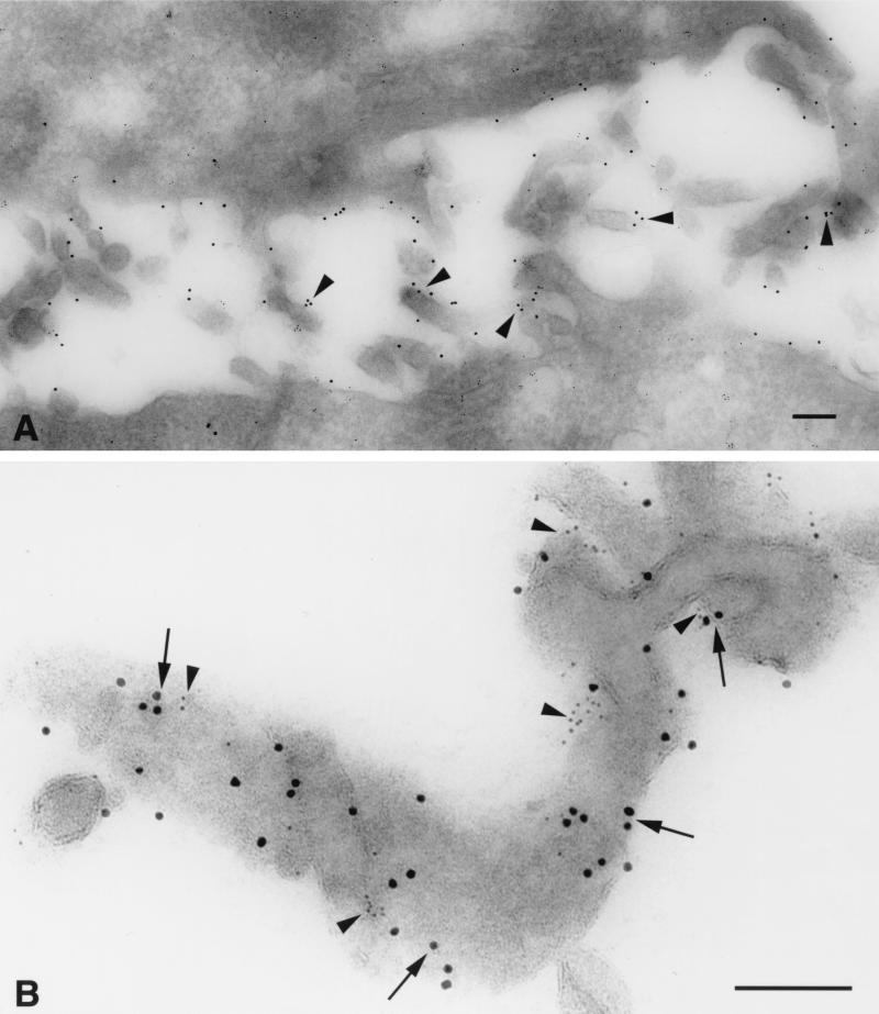
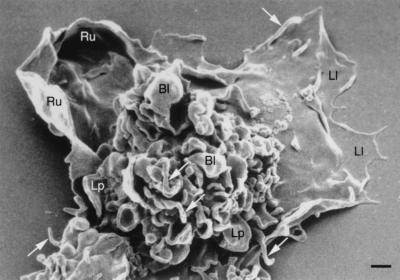
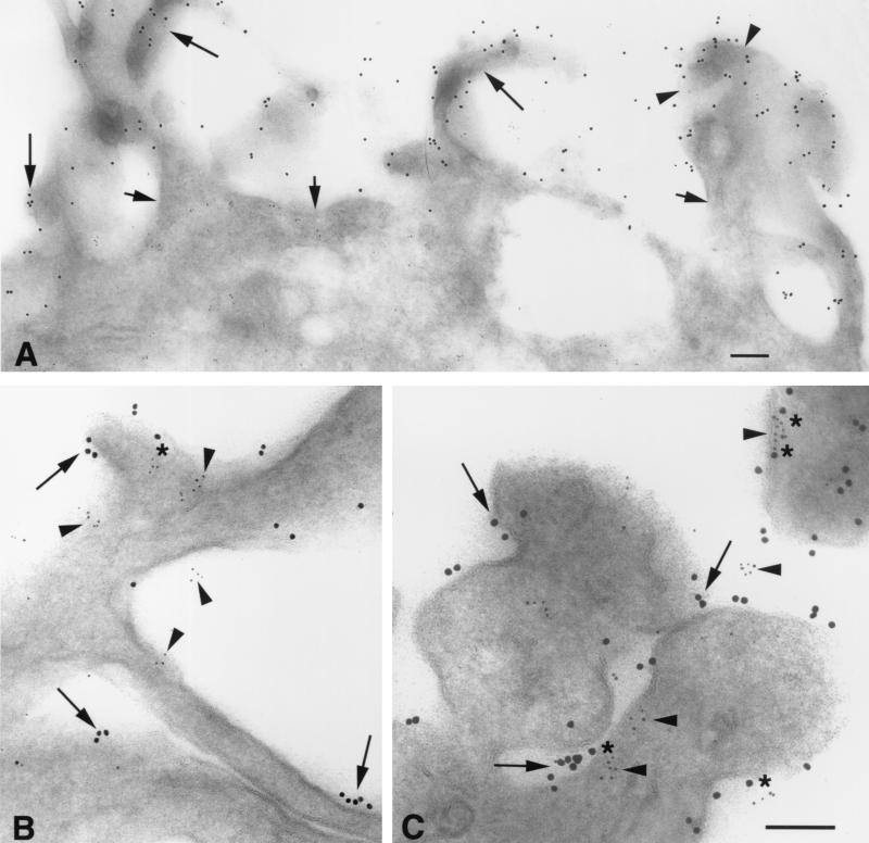
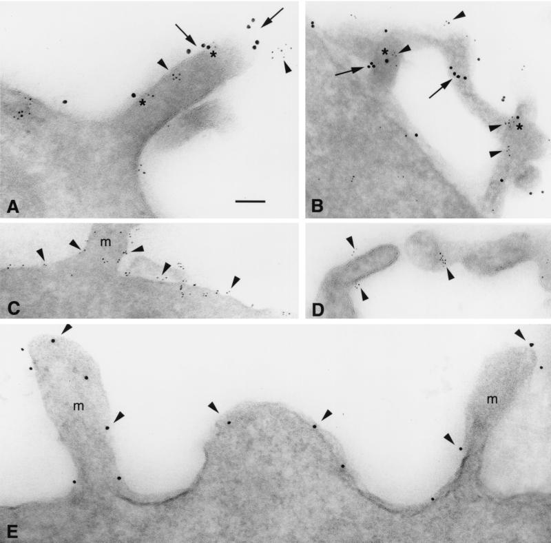
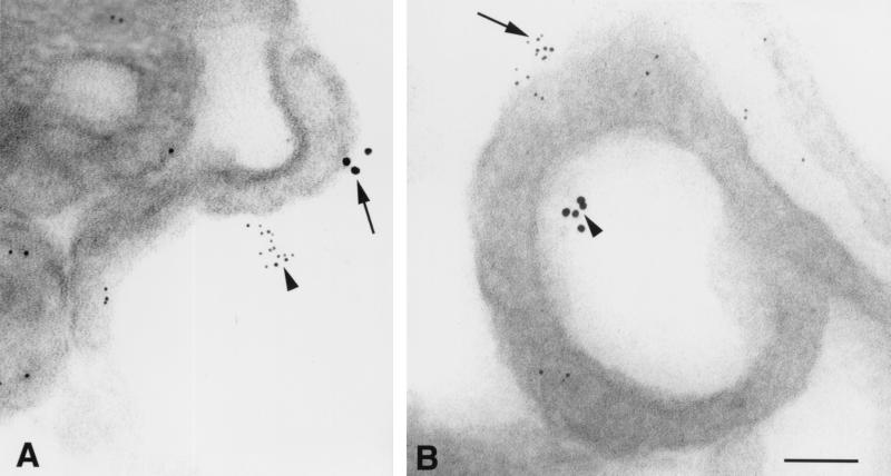
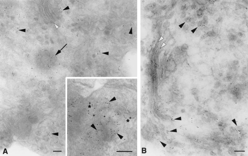
The chemokine receptors CCR5 and CXCR4 act synergistically with CD4 in an ordered multistep mechanism to allow the binding and entry of human immunodeficiency virus type 1 (HIV-1). The efficiency of such a coordinated mechanism depends on the spatial distribution of the participating molecules on the cell surface. Immunoelectron microscopy was performed to address the subcellular localization of the chemokine receptors and CD4 at high resolution. Cells were fixed, cryoprocessed, and frozen; 80-nm cryosections were double labeled with combinations of CCR5, CXCR4, and CD4 antibodies and then stained with immunogold. Surprisingly, CCR5, CXCR4, and CD4 were found predominantly on microvilli and appeared to form homogeneous microclusters in all cell types examined, including macrophages and T cells. Further, while mixed microclusters were not observed, homogeneous microclusters of CD4 and the chemokine receptors were frequently separated by distances less than the diameter of an HIV-1 virion. Such distributions are likely to facilitate cooperative interactions with HIV-1 during virus adsorption to and penetration of human leukocytes and have significant implications for development of therapeutically useful inhibitors of the entry process. Although the mechanism underlying clustering is not understood, clusters were observed in small trans-Golgi vesicles, implying that they were organized shortly after synthesis and well before insertion into the cellular membrane. Chemokine receptors normally act as sensors, detecting concentration gradients of their ligands and thus providing directional information for cellular migration during both normal homeostasis and inflammatory responses. Localization of these sensors on the microvilli should enable more precise monitoring of their environment, improving efficiency of the chemotactic process. Moreover, since selectins, some integrins, and actin are also located on or in the microvillus, this organelle has many of the major elements required for chemotaxis.


Figures

FIG. 1
FIG. 1
Δ10 CCR5 has greatly diminished affinity for gp120. The avidity of R5-tropic gp120s, JRFL (◊) or YU2 (▵), for the target receptor was measured by their ability to compete against 125I-MIP-1α in the presence of soluble CD4. Experiments were carried out with CHO cells expressing either wild-type CCR5 (open symbols) or Δ10 CCR5 (filled symbols).
FIG. 2
FIG. 2
Western blots demonstrate that each chemokine receptor IgG used for immuno-EM recognizes only the receptor from which its immunizing peptide was derived. Specificities were tested against extracts of whole cells expressing either hCCR1 (lane 1), hCCR2 (lane 2), hCCR3 (lane 3), hCXCR4 (lane 4), or hCCR5 (lane 5). Results are for the N-terminal CCR5 (R4603; A), C-terminal CCR5 (R4627; B), N-terminal CXCR4 (R5039; C), and N-terminal CCR2 (R4731; D) antibodies. Sizes are indicated in kilodaltons.
FIG. 3
FIG. 3
Double-label immunofluorescence microscopy shows that CCR5 (A, C, and E) and CD4 (B, D, and F) are colocalized on the surfaces of HeLa-C29 cells (A and B), human macrophages (C and D), and human T cells (E and F) (bar = 20 μm). CCR5 was frequently found to be coincident with CD4 at the leading edge of human macrophages (arrowheads in panels C and D).
FIG. 4
FIG. 4
Immuno-EM detects homogeneous microclusters of CCR5 and CD4 concentrated on the microvilli of HeLa-C29 cells. (A) CCR5 is preferentially localized on cell surface microvilli (arrowheads; 10-nm anti-rabbit immunogold). (B) Prominent homogeneous microclusters of CCR5 (arrows; 10-nm anti-rabbit immunogold) and CD4 (arrowheads; 5-nm anti-mouse immunogold) are closely apposed on the surface membranes of microvilli. The central arrowhead depicts a linear array of CD4 epitopes situated within 10 nm of a CCR5 immunogold particle in the outer membrane glycocalyx. Bars = 100 nm.
FIG. 5
FIG. 5
Scanning electron micrograph of a typical cultured human blood macrophage. The cells exhibit structures reflecting intense surface membrane activity: prominent microvilli (white arrows), blebs (Bl), lamellipodia (Lp), ruffling membranes (Ru), and leading lamellae (Ll). Bar = 1 μm.
FIG. 6
FIG. 6
CCR5 and CD4 form homogeneous microclusters on microvilli of human blood macrophages detected by immuno-EM. (A) CD4 (10-nm immunogold) is concentrated on microvilli (long arrows) and blebs (arrowheads), while little staining is apparent on the cell surface membrane (short arrows). Ultrathin cryosections through microvilli (B) and blebs (C) exhibit homogeneous microclusters of CCR5 (arrowheads; 5-nm immunogold) and CD4 (arrows; 10-nm immunogold) localized on their surface membranes; asterisks indicate closely apposed CCR5 and CD4. A complex of CCR5 and CD4 (C, upper right corner) contains two loci of CD4 epitopes (asterisks) closely flanking an elongated CCR5 aggregate on the cell membrane (arrowhead). Bars = 100 nm.
FIG. 7
FIG. 7
Immuno-EM exhibits homogeneous microclusters of CCR5, CXCR4, and CD4 on primary human T cells. (A) T-cell microvilli exhibit homogeneous microaggregates of CCR5 (arrowheads; 5-nm immunogold) and CD4 (arrows; 10-nm immunogold); asterisks indicate closely apposed CCR5 and CD4 epitopes. (B) CXCR4 (arrowheads; 5-nm immunogold) exhibits similar homogeneous microclusters, closely apposed (at asterisks) to CD4 (arrows; 10-nm immunogold) on T-cell microvilli. (C) CD3 (arrowheads) is localized on T-cell microvilli (m) and over the entire cell surface membrane. (D) CD8 preferentially labels T-cell microvilli as small aggregates (arrowheads) and is not detected on the surface membrane. (E) gp120 epitopes (arrowheads; 10-nm immunogold) appear unclustered and randomly distributed on microvilli (m) and the cell membranes of CHO cells expressing 105 YU2 gp143 copies per cell (labeled with 1b12, a human MAb to gp120). Bar (applies to all panels) = 100 nm.
FIG. 8
FIG. 8
CCR5 and CXCR4 are localized in separate microclusters on human macrophages. (A) Arrowhead shows a homogeneous microcluster of CXCR4 stained with an N-terminal rabbit anti-peptide IgG, and the arrow depicts a separate microaggregate of CCR5 labeled with MAb 2D7; both clusters are localized on a single microvillus. (B) A CCR5 microaggregate is labeled with a C-terminal rabbit antipeptide IgG (arrow), while a CXCR4 microcluster is stained with MAb 12G5 (arrowhead); both are located on the same lamellipodium. Bar = 100 nm.
FIG. 9
FIG. 9
Microclusters of CCR5 are localized in Golgi secretory vesicles in human macrophages and HeLa-C29 cells. (A) Cryosection through the Golgi apparatus of a human macrophage labeled for CCR5 with 5-nm immunogold. Microaggregates of CCR5 epitopes are localized as clusters (arrowheads) or curvilinear arrays (arrow) in Golgi secretory vesicles. Little CCR5 label is present in the Golgi cysternae (white arrowhead). (Inset) Grazing section through membrane blebs at the macrophage surface double labeled for CCR5 and CD4. Curvilinear aggregates of CCR5 epitopes (arrowheads; 5-nm immunogold) are associated with a dense submembranous cortex and with CD4 labeling (asterisks; 10-nm immunogold). This image suggests that CCR5-positive secretory vesicles are fusing with the cell membrane. (B) Localization of CCR5 (5-nm immunogold) in the Golgi apparatus of a HeLa-C29 cell. Microclusters of CCR5 are concentrated in Golgi secretory vesicles (arrowheads), while reduced CCR5 labeling is detected in the Golgi cysternae (white arrowheads). Bars = 100 nm.

Similar articles

Cited by

References

    1. Aithal H N, Knigge K M, Kartha S, Czyzewski E A, Toback F G. An alternate method utilizing small quantities of ligand for affinity purification of monospecific antibodies. J Immunol Methods. 1988;112:63–69. - PubMed
    1. Alfano M, Schmidtmayerova H, Amella C A, Pushkarsky T, Bukrinsky M. The B-oligomer of pertussis toxin deactivates CC chemokine receptor 5 and blocks entry of M-tropic HIV-1 strains. J Exp Med. 1999;190:597–605. - PMC - PubMed
    1. Alkhatib G, Combadiere C, Broder C C, Feng Y, Kennedy P E, Murphy P M, Berger E A. CC CKR5: a RANTES, MIP-1α, MIP-1β receptor as a fusion cofactor for macrophage-tropic HIV-1. Science. 1996;272:1955–1958. - PubMed
    1. Baba M, Nishimura O, Kanzaki N, Okamoto M, Sawada H, Iizawa Y, Shiraishi M, Aramaki Y, Okonogi K, Ogawa Y, Meguro K, Fujino M. A small-molecule, nonpeptide CCR5 antagonist with highly potent and selective anti-HIV-1 activity. Proc Natl Acad Sci USA. 1999;96:5698–5703. - PMC - PubMed
    1. Baggiolini M, Dewald B, Moser B. Human chemokines: an update. Annu Rev Immunol. 1997;15:675–705. - PubMed

MeSH terms