. 2001 Apr;75(8):3832-40.
doi: 10.1128/JVI.75.8.3832-3840.2001.

Requirements for the nuclear-cytoplasmic translocation of infected-cell protein 0 of herpes simplex virus 1

Affiliations

Requirements for the nuclear-cytoplasmic translocation of infected-cell protein 0 of herpes simplex virus 1

P Lopez et al. J Virol. 2001 Apr.

Abstract

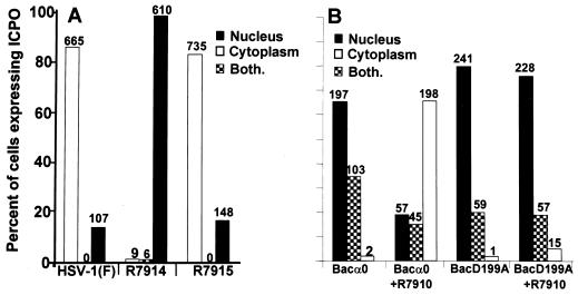
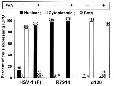
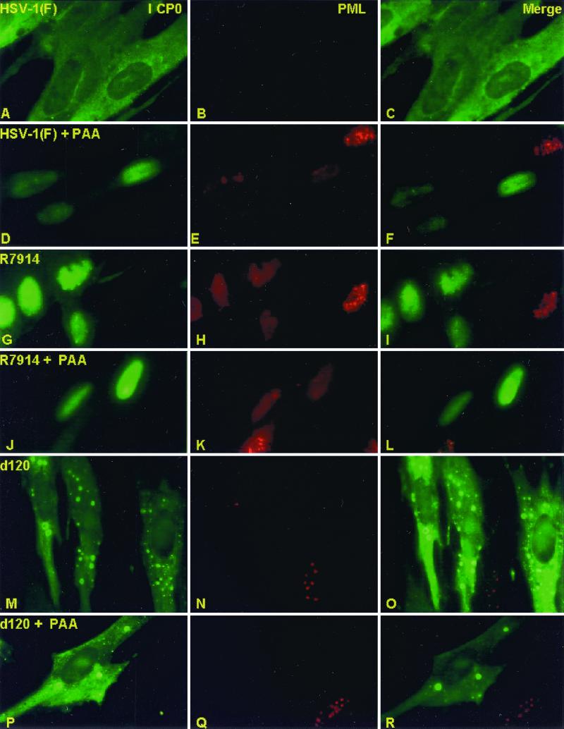
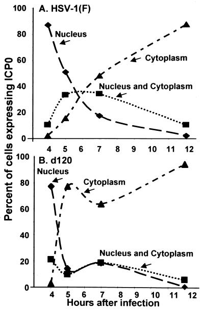
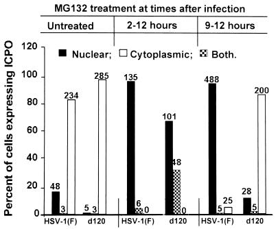
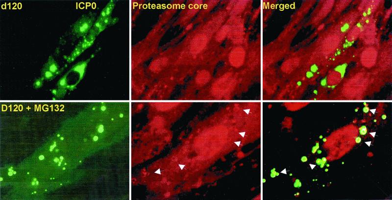
Earlier studies have shown that wild-type infected-cell protein 0 (ICP0), a key herpes simplex virus regulatory protein, translocates from the nucleus to the cytoplasm of human embryonic lung (HEL) fibroblasts within several hours after infection (Y. Kawaguchi, R. Bruni, and B. Roizman, J. Virol. 71:1019-1024, 1997). Translocation of ICP0 was also observed in cells infected with the d120 mutant, in which both copies of the gene encoding ICP4, the major regulatory protein, had been deleted (V. Galvan, R. Brandimarti, J. Munger, and B. Roizman, J. Virol. 74:1931-1938, 2000). Furthermore, a mutant (R7914) carrying the D199A substitution in ICP0 does not bind or stabilize cyclin D3 and is retained in the nucleus (C. Van Sant, P. Lopez, S. J. Advani, and B. Roizman, J. Virol. 75:1888-1898, 2001). Studies designed to elucidate the requirements for the translocation of ICP0 between cellular compartments revealed the following. (i) Translocation of ICP0 to the cytoplasm in productive infection maps to the D199 amino acid, inasmuch as wild-type ICP0 delivered in trans to cells infected with an ICP0 null mutant was translocated to the cytoplasm whereas the D199A-substituted mutant ICP0 was not. (ii) Translocation of wild-type ICP0 requires a function expressed late in infection, inasmuch as phosphonoacetate blocked the translocation of ICP0 in wild-type virus-infected cells but not in d120 mutant-infected cells. Moreover, whereas in d120 mutant-infected cells ICP0 was translocated rapidly from the cytoplasm to the nucleus at approximately 5 h after infection, the translocation of ICP0 in wild-type virus-infected cells extended from 5 to at least 9 h after infection. (iii) In wild-type virus-infected cells, the MG132 proteasomal inhibitor blocked the translocation of ICP0 to the cytoplasm early in infection, but when added late in infection, it caused ICP0 to be relocated back to the nucleus from the cytoplasm. (iv) MG132 blocked the translocation of ICP0 in d120 mutant-infected cells early in infection but had no effect on the ICP0 aggregated in vesicle-like structures late in infection. However, in d120 mutant-infected cells treated with MG132 at late times, proteasomes formed a shell-like structure around the aggregated ICP0. These structures were not seen in wild-type virus or R7914 mutant-infected cells. The results indicate the following. (i) In the absence of beta or gamma protein synthesis, ICP0 dynamically associates with proteasomes and is translocated to the cytoplasm. (ii) In cells productively infected beyond alpha gene expression, ICP0 is retained in the nucleus until after the onset of viral DNA synthesis and the synthesis of gamma2 proteins. (iii) Late in infection, ICP0 is actively sequestered in the cytoplasm by a process mediated by proteasomes, inasmuch as interference with proteasomal function causes rapid relocation of ICP0 to the nucleus.


Figures

FIG. 1
FIG. 1
The requirements for the translocation of ICP0 from the nucleus to the cytoplasm. (A) ICP0 carrying the D199A substitution encoded by R7914 is not translocated to the cytoplasm late in infection of HEL fibroblasts. Replicate slide cultures of HEL fibroblasts were infected with 20 PFU of HSV-1(F), the recombinant virus R7914, or the repaired virus R7915 per cell and maintained at 37°C. At 12 h after infection, the cells were fixed and reacted first with polyclonal rabbit serum directed against exon II of ICP0 and second with FITC-conjugated goat anti-rabbit immunoglobulin antibodies. Sequential fields were examined in a Zeiss confocal microscope, and the numbers of cells exhibiting nuclear, cytoplasmic, or both nuclear and cytoplasmic localization of ICP0 were tabulated as shown in the histogram. The numbers above the bars indicate the numbers of cells showing a specific distribution of ICP0. (B) A viral gene function mediates the translocation of ICP0 from the nucleus to the cytoplasm during productive infection. Replicate slide cultures of HEL fibroblasts were exposed first to recombinant baculoviruses encoding wild-type or mutant ICP0 (D199A). After 2 h, cells were treated with 5 mM Na-butyrate. At 3 h after exposure to baculoviruses, the cells were exposed to the recombinant virus R7910, from which both copies of the α0 gene had been deleted. After 15 h of exposure to baculoviruses alone or 12 h after infection with R7910, the cells were fixed and reacted with rabbit polyclonal antibody against ICP0 and mouse monoclonal antibody against gD and then reacted with FITC-conjugated goat anti-rabbit IgG and Texas Red-conjugated goat anti-mouse IgG antibodies. Cell counts were done as described above, except that only cells exhibiting gD encoded by R7910 and ICP0 encoded by baculoviruses were counted.
FIG. 2
FIG. 2
Translocation of ICP0 from the nucleus to the cytoplasm in HEL fibroblasts infected with wild-type virus is blocked by PAA. Replicate slide cultures of HEL fibroblasts were infected with 20 PFU of HSV-1(F), R7914, or d120 mutant virus per cell in the presence or absence of 300 μg of PAA per ml of medium. The cells were fixed at 12 h after infection and reacted with polyclonal rabbit antibody against ICP0 and then with FITC-conjugated goat anti-rabbit IgG. The tabulation of cells exhibiting ICP0 in the nucleus, the cytoplasm, or both was done as described in the legend to Fig. 1.
FIG. 3
FIG. 3
Immunofluorescent images of HEL fibroblasts infected with HSV-1(F), R7914, or d120 mutant and either treated or not treated with PAA. The images shown are representative of infected cells treated, generated, and counted as described in the legend to Fig. 2. The cells were reacted with rabbit polyclonal antibody to ICP0 and mouse monoclonal antibody to PML and then reacted with goat anti-rabbit IgG conjugated to FITC and goat anti-mouse IgG conjugated to Texas Red. The left, middle, and right columns show the cell localization of ICP0 and PML and merged images, respectively. The images were captured with a Zeiss confocal microscope with the aid of software provided by the manufacturer.
FIG. 4
FIG. 4
Temporal pattern of translocation of ICP0 encoded by wild-type and d120 mutant viruses from the nucleus to the cytoplasm. Replicate slide cultures of HEL fibroblasts were exposed to 20 PFU of HSV-1(F) or d120 mutant virus per cell. At 4, 5, 7, or 12 h after infection and incubation at 37°C, replicate cultures were fixed and reacted with rabbit polyclonal antibody against ICP0 and then with goat anti-rabbit IgG conjugated to FITC. The cells were examined and tabulated as described in the legend to Fig. 1.
FIG. 5
FIG. 5
Effect of MG132 proteasomal inhibitor on the translocation of ICP0 from the nucleus to the cytoplasm. Replicate slide cultures of HEL fibroblasts were exposed to 20 PFU of HSV-1(F) or d120 mutant virus per cell. At 2 h after infection (middle bars) or 9 h after infection (right bars), the cells were replenished with medium containing MG132 (5 μM). At 12 h after infection, the cells were fixed and reacted with polyclonal rabbit antibody against ICP0 and then with goat anti-rabbit IgG conjugated to FITC. The cells were examined and quantified as described in the legend to Fig. 1.
FIG. 6
FIG. 6
Colocalization of proteasome and ICP0 in cells infected with the d120 mutant and exposed to MG132. The experimental design of this series of experiments was identical to that described in the legend to Fig. 3, except that the cells were reacted with mouse monoclonal antibody against ICP0 and rabbit polyclonal antibody to the whole proteasome designated as core antibody (Affiniti Research Products Ltd.). The secondary antibodies were goat anti-mouse IgG conjugated to FITC and goat anti-rabbit IgG conjugated to Texas Red. Shown are images of cells infected with d120 mutant virus and either left untreated (upper panels) or exposed at 9 h after infection to 5 μM MG132 (lower panels). The images were captured with a Zeiss confocal microscope with the aid of software provided by the manufacturer. The arrows point to proteasomal core proteins aggregated in the form of a shell surrounding ICP0 in the cytoplasm of MG132-treated cells. These structures were seen only in d120 mutant-infected cells treated with MG132.

Similar articles

Cited by

References

    1. Blaho J A, Mitchell C, Roizman B. An amino acid sequence shared by the herpes simplex virus 1 alpha regulatory proteins 0, 4, 22, and 27 predicts the nucleotidylylation of the UL21, UL31, UL47, and UL49 gene products. J Biol Chem. 1994;269:17401–17410. - PubMed
    1. Cai W Z, Schaffer P A. Herpes simplex virus type 1 ICP0 plays a critical role in the de novo synthesis of infectious virus following transfection of viral DNA. J Virol. 1989;63:4579–4589. - PMC - PubMed
    1. Chelbi-Alix K M, de The H. Herpes virus induced proteasome-dependent degradation of the nuclear bodies-associated PML and Sp100 proteins. Oncogene. 1999;18:935–941. - PubMed
    1. DeLuca N A, McCarthy A M, Schaffer P A. Isolation and characterization of deletion mutants of herpes simplex virus type 1 in the gene encoding immediate-early regulatory protein ICP4. J Virol. 1985;56:558–570. - PMC - PubMed
    1. Ejercito P M, Kieff E D, Roizman B. Characterization of herpes simplex virus strains differing in their effects on social behaviour of infected cells. J Gen Virol. 1968;2:357–364. - PubMed

Publication types

MeSH terms

LinkOut - more resources