. 2001 Apr;75(8):3885-95.
doi: 10.1128/JVI.75.8.3885-3895.2001.

Herpes simplex virus type 1 promoter activity during latency establishment, maintenance, and reactivation in primary dorsal root neurons in vitro

Affiliations

Herpes simplex virus type 1 promoter activity during latency establishment, maintenance, and reactivation in primary dorsal root neurons in vitro

J L Arthur et al. J Virol. 2001 Apr.

Abstract

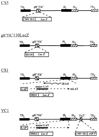
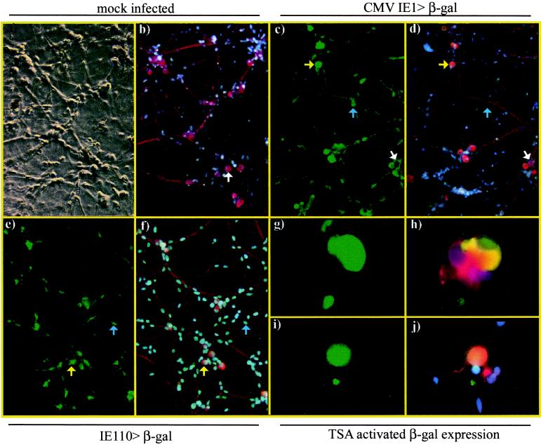
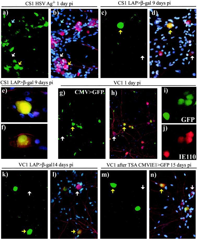
A neonatal rat dorsal root ganglion-derived neuronal culture system has been utilized to study herpes simplex virus (HSV) latency establishment, maintenance, and reactivation. We present our initial characterization of viral gene expression in neurons following infection with replication-defective HSV recombinants carrying beta-galactosidase and/or green fluorescent protein reporter genes under the control of lytic cycle- or latency-associated promoters. In this system lytic virus reporter promoter activity was detected in up to 58% of neurons 24 h after infection. Lytic cycle reporter promoters were shut down over time, and long-term survival of neurons harboring latent virus genomes was demonstrated. Latency-associated promoter-driven reporter gene expression was detected in neurons from early times postinfection and was stably maintained in up to 83% of neurons for at least 3 weeks. In latently infected cultures, silent lytic cycle promoters could be activated in up to 53% of neurons by nerve growth factor withdrawal or through inhibition of histone deacetylases by trichostatin A. We conclude that the use of recombinant viruses containing reporter genes, under the regulation of lytic and latency promoter control in neuronal cultures in which latency can be established and reactivation can be induced, is a potentially powerful system in which to study the molecular events that occur during HSV infection of neurons.


Figures

FIG. 1
FIG. 1
Schematic of gHTK recombinant viruses. All viruses were made gHTK through the deletion of nucleotides 43771 to 47103 spanning the gH and TK coding sequences. Virus CS5 contains a CMV IE1 promoter-driven lacZ expression cassette replacing gH and TK sequences. Virus gHTK110LacZ contains an IE110 promoter-driven lacZ expression cassette inserted at the same locus. Virus CS1 contains an EMCV IRES-linked lacZ gene inserted between the HpaI sites within major LATs and was generated by the deletion of gH and TK from SC16 LβA. Virus VC1 is derived from coinfection of CS1 and C12 and consists of the CMV IE1 promoter-driven EGFP reporter gene cassette in the Us5 locus from C12 on the CS1 backbone. In VC1 and CS1, β-gal expression is driven by the endogenous LAP.
FIG. 2
FIG. 2
Representative examples of mock-infected neuronal cultures or cultures infected with 106 PFU of CS5 (CMV IE1>β-gal) or gHTK110LacZ (IE110>β-gal) per well. Cultures were fixed and dually immunostained for expression of β-gal (FITC, green) and neuron-specific β-tub (Cy3, red) and were counterstained with DAPI to show all cell nuclei (blue). An example is given of mock-infected cultures showing identification of neurons by β-tub staining (a and b, arrows). Examples are given of β-gal expression at day 1 p.i. in neurons (i.e., yellow arrows) and nonneuronal cells (i.e., blue arrows) in wells infected with CS5 (c, d) and gHTK110LacZ (e, f). An example of β-gal-negative neurons is indicated (white arrows). Examples are given of β-gal expression in CS5-infected (g, h) and gHTK110LacZ-infected (i, j) cultures at 15 days p.i., 24 h after the addition of 660 nM TSA. The small areas of β-gal fluorescence do not have nuclei and likely indicate antibody binding to cellular debris. Digital photomicrographs were taken as phase-contrast (a) or fluorescence images using either the FITC filter for β-gal (c, e, g, i) or the triple band-pass filter (b, d, f, h, j) to allow covisualization of FITC, Cy3, and DAPI fluorescence in which colocalization of β-gal and β-tub gives a yellow-orange signal.
FIG. 3
FIG. 3
Representative examples of neuronal cultures infected with 5 × 10⁵ PFU of either CS1 (a to f) or VC1 (g, h, j to m) per well or 3 × 10⁵ PFU of VC1 (i, j) per well. Fixed cultures were stained by dual (a to f, k, l) or single (g to k, m, n) immunofluorescence to detect expression of viral gene products (green; also see panel h) and/or β-tub (red). Cultures were counterstained with DAPI to show cell nuclei (blue) and were visualized via fluorescence by using either the FITC filter to show FITC (a, c, k) and GFP (g, i, m) or the triple band-pass filter to allow covisualization of DAPI, Cy3 with FITC (b, d to f, k), or GFP signals (h, m). Examples indicated are HSV Ag+, GFP+, or β-gal+ neurons (yellow arrows); HSV Ag+ nonneuronal cells (blue arrows); or neurons in which viral gene products are not detected (white arrows). At day 1 p.i., HSV Ag expression from CS1 (a, b) and CMV IE1 promoter-driven GFP expression from VC1 (k, l) or IE110 (i, j) were detected in neurons and nonneuronal cells. LAP-driven β-gal expression was detected in a proportion of neurons 9 days p.i. with CS1 (c to f) or 14 days p.i. with VC1 (k, l), and higher magnifications show examples of the intense (e) or average (f) β-gal staining obtained. Addition of 660 nM TSA to VC1-infected cultures 14 days p.i. resulted in detection of GFP expression in some neurons 24 h later (m, n).

References

    1. Alvira R M, Goins W F, Cohen J B, Glorioso J G. Genetic studies exposing the splicing events involved in herpes simplex virus type 1 latency-associated transcript production during lytic and latent infection. J Virol. 1999;73:3866–3878. - PMC - PubMed
    1. Arthur J, Efstathiou S, Simmons A. Intranuclear foci containing low abundance herpes simplex virus latency-associated transcripts visualized by non-isotopic in situ hybridization. J Gen Virol. 1993;74:1363–1370. - PubMed
    1. Arthur J L, Everett R, Brierley I, Efstathiou S. Disruption of the 5′ and 3′ splice sites flanking the 2-kilobase latency-associated transcripts: evidence for alternate splicing in lytic and latent infections. J Gen Virol. 1998;79:107–116. - PubMed
    1. Babic N, Rodger G, Arthur J, Minson A C. A study of primary neuronal infection by mutants of herpes simplex virus type 1 lacking dispensable and non-dispensable glycoproteins. J Gen Virol. 1999;80:2403–2409. - PubMed
    1. Block T M, Spivack J G, Steiner I, Deshmane S, McIntosh M T, Lirette R P, Fraser N W. A herpes simplex virus type 1 latency-associated transcript mutant reactivates with normal kinetics from latent infection. J Virol. 1990;64:3417–3426. - PMC - PubMed

Publication types

MeSH terms

LinkOut - more resources