

doi: 10.1128/JVI.75.8.4002-4007.2001.
Biophysical characterization and vector-specific antagonist activity of domain III of the tick-borne flavivirus envelope protein
Affiliations
- PMID: 11264392
- PMCID: PMC114894
- DOI: 10.1128/JVI.75.8.4002-4007.2001
Item in Clipboard
Biophysical characterization and vector-specific antagonist activity of domain III of the tick-borne flavivirus envelope protein
J Virol.
2001 Apr.
Abstract
The molecular determinants responsible for flavivirus host cell binding and tissue tropism are largely unknown, although domain III of the envelope protein has been implicated in these functions. We examined the solution properties and antagonist activity of Langat virus domain III. Our results suggest that domain III adopts a stably folded structure that can mediate binding of tick-borne flaviviruses but not mosquito-borne flaviviruses to their target cells. Three clusters of phylogenetically conserved residues are identified that may be responsible for the vector-specific antagonist activity of domain III.
Figures
Coomassie blue-stained composite sodium dodecyl sulfate-polyacrylamide gel showing expression and purification of recombinant LgtE-D3. Lane 1, GST–LgtE-D3 fusion protein bound to glutathione-conjugated agarose beads. Lane 2, total protein at the initiation of thrombin cleavage of bound GST–LgtE-D3. Lane 3, soluble protein after thrombin cleavage of bound GST–LgtE-D3. Lanes 4 to 6, soluble protein recovered after sequential washes of glutathione-agarose beads with low-salt buffer. Lane 7, protein bound to glutathione-agarose beads after thrombin cleavage. Lane 8, Western immunoblot of purified LgtE-D3 probed with rabbit polyclonal antibody generated against recombinant LgtE-D3 (Alpha Diagnostics, Inc.). The GST–LgtE-D3 fusion protein migrates as a single band at ∼37 kDa. Recombinant LgtE-D3 migrates as a single band at ∼10.7 kDa.
Far-UV CD spectra of recombinant LgtE-D3 incubated with increasing amounts of denaturant. Spectra from LgtE-D3 in 0 M urea (solid curve, solid circles), 2 M urea (dotted curve, open circles), 4 M urea (dotted curve, open squares), and 8 M urea (dashed curve, open triangles) are shown. The LgtE-D3 concentration was 0.3 mg/ml in Tris buffer (pH 7.4). Spectra were collected on an Aviv 62 DS circular dichrometer operating with a 0.5-nm step increment and a 1-s interval. Cylindrical quartz cuvettes with a 0.1-cm path length were used for all measurements. All sample spectra were recorded five times, averaged, and corrected for buffer contributions. Measurements were considered unreliable when the instrument dynode voltage exceeded 410 V and were not included in subsequent analyses.
Amino acid sequence alignment of domain III of flavivirus E proteins. Residues that are conserved in at least 15 of the 16 sequences are indicated by a star above the alignment. Shaded boxes indicate positions of conserved vector-specific residues and insertions. Amino acid numbering refers to TBE virus residue positions. The virus and vector classes are indicated along the left-hand margin. Protein sequences were obtained from Swiss-Protein database entries and correspond to Langat virus (LGT; strain V, accession number P29837), Powassan virus (POW; accession number Q04538), Central European TBE virus (accession number Q01299), louping ill virus (LI; accession number P22338), yellow fever virus (YF; strain 1899/81, accession number P29165), Murray Valley encephalitis virus (MV; accession number P05769), Japanese encephalitis virus (JE; strain Nakayama, accession number P27395), St. Louis encephalitis virus (SLE; accession number P09732, strain MS1-7), Kunjin virus (KUN; strain MRM61C, accession number P14335), West Nile virus (WN; strain RO97-50, accession number Q9WHD1), dengue virus type 1 (DEN1; strain AHF82-80, accession number P27912), dengue virus type 2 (DEN2; isolate Malaysia M2, accession number P14338), dengue virus type 3 (DEN3; accession number P27915), dengue virus type 4 (DEN4; accession number P09866), Rio Bravo virus (RIO; accession number AF144692), and Apoi virus (accession number AF160193).
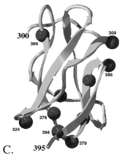
Structure of TBE virus envelope protein. (A) Top view of the E protein homodimer. (B) Side view of the E protein homodimer. Orientation nomenclature is adapted from that of Rey et al. (13), where top suggests a view towards and normal to the virus surface, and side suggests a view tangent to the virus surface, with the viral membrane below the protein. Each monomer of the homodimer is shaded differently, and the dashed lines delineate the domain III boundaries. (C) Isolated domain III viewed approximately perpendicular to the twofold axis of the E protein homodimer, corresponding to the solvent-exposed lateral surface of domain III. Positions of conserved vector-specific residues and insertions in domain III are indicated with spheres. Amino acid numbering refers to the TBE virus residue positions. Domain III structures were displayed using Swiss-Pdb Viewer (5; http://www.expasy.ch/spdbv ).
Structure of TBE virus envelope protein. (A) Top view of the E protein homodimer. (B) Side view of the E protein homodimer. Orientation nomenclature is adapted from that of Rey et al. (13), where top suggests a view towards and normal to the virus surface, and side suggests a view tangent to the virus surface, with the viral membrane below the protein. Each monomer of the homodimer is shaded differently, and the dashed lines delineate the domain III boundaries. (C) Isolated domain III viewed approximately perpendicular to the twofold axis of the E protein homodimer, corresponding to the solvent-exposed lateral surface of domain III. Positions of conserved vector-specific residues and insertions in domain III are indicated with spheres. Amino acid numbering refers to the TBE virus residue positions. Domain III structures were displayed using Swiss-Pdb Viewer (5; http://www.expasy.ch/spdbv ).
References
-
- Barrett A D T. Japanese encephalitis and dengue vaccines. Biologicals. 1997;25:27–34. - PubMed
-
- Barrett A D T. Yellow fever vaccines. Biologicals. 1997;25:17–25. - PubMed
-
- Chen Y, Maquire T, Hileman R E, Fromm J R, Esko J D, Linhardt R J, Marks R M. Dengue virus infectivity depends on envelope protein binding to target cell heparan sulfate. Nat Med. 1997;3:866–871. - PubMed
-
- Dunster L M, Wang H, Ryman K D, Miller B R, Watowich S J, Minor P D, Barrett A D T. Molecular and biological changes associated with HeLa cell attenuation of wild-type yellow fever virus. Virology. 1999;261:309–318. - PubMed
-
- Guex N, Peitsch M C. SWISS-MODEL and the Swiss-Pdb Viewer: an environment for comparative protein modeling. Electrophoresis. 1997;18:2714–2723. - PubMed
Publication types
MeSH terms
Substances
LinkOut - more resources
Full Text Sources
Other Literature Sources
