. 2000 Aug;1(2):145-50.
doi: 10.1093/embo-reports/kvd023.

Fate of mat1 DNA strands during mating-type switching in fission yeast

Affiliations

Fate of mat1 DNA strands during mating-type switching in fission yeast

B Arcangioli. EMBO Rep. 2000 Aug.

Abstract

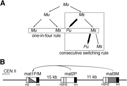
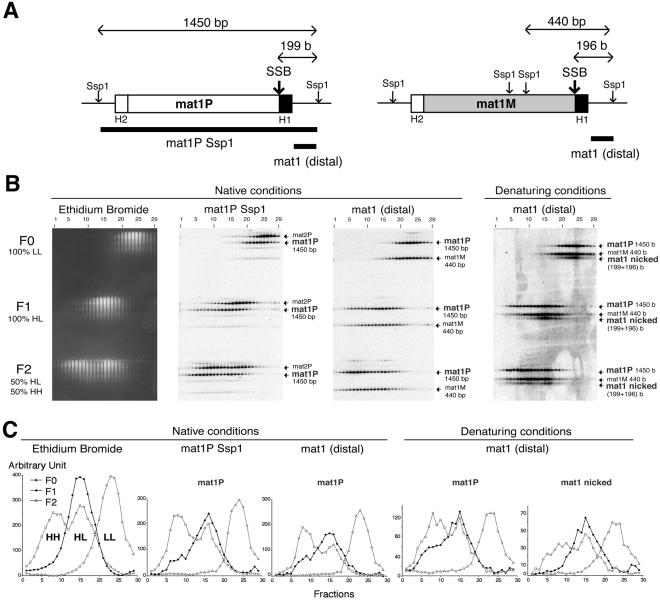
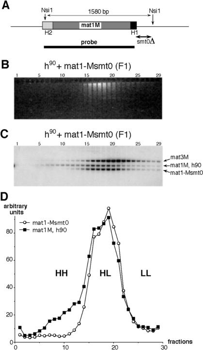
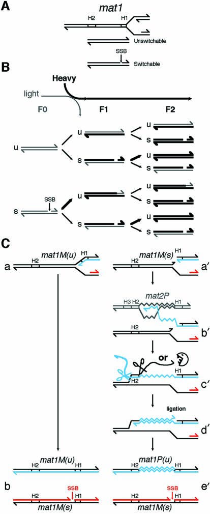
The mating-type switching of the fission yeast, Schizosaccharomyces pombe, is highly regulated. Two consecutive asymmetric divisions are required to produce one mating-type switched cell among the four progeny. Using DNA density-gradient centrifugation we demonstrate that one-fourth of the mat1 DNA is not replicated by the conventional semi-conservative mode, but instead both DNA strands are synthesized de novo. Our data are consistent with a gene conversion event, initiated by a site- and strand-specific DNA break (SSB). We further demonstrate that the virgin switched mat1-containing chromatid no longer contained the nick, while it is reintroduced during the lagging strand synthesis of the mat1 locus on the sister chromatid. This finding establishes at the molecular level a firm experimental link between the phenotype and genotype in the process of asymmetric mating-type switching during mitotic divisions.


Figures

None
Fig. 1. Mating-type pedigree and loci of fission yeast. (A) P and M indicate the mating-type of the cell, the suffix u or s represents the unswitchable and switchable potential of the cell, respectively. For simplicity, only the M lineage is shown. The one-in-four and the consecutive switching rules are indicated. (B) The mating-type region on chromosome II. The mat1 locus contains either the P (white box) or the M (grey box) alleles, and mat2P and mat3M are donors of genetic information. The H1 and H2 homology sequences are common to all cassettes whereas the H3 sequence is common to only the silent mat2P and mat3M loci (Kelly et al., 1988).
None
Fig. 2. Density distribution of the mat1 locus. (A) The location of the SspI restriction sites at the mat1 locus and their respective sizes are indicated. The position of the site- and strand-specific break (SSB) is shown by an arrow. 199 and 196 bases are the sizes of the mat1P- and mat1M-distal upper DNA strand, following SspI digestion. The H1 and H2 sequences are indicated and the two DNA probes used are shown at the bottom. (B) Distribution of the mat1 locus analysed by DNA–DNA hybridization. DNA samples from the CsCl-eluted fractions were electrophoresed in native or denaturing conditions, as indicated. F0, F1, F2, LL, HL and HH indicate the number of generations in heavy media and the double-stranded DNA density, respectively. The probes used (A) are double stranded and are indicated at the top of the autoradiogram. Note that the same probe, mat1 (distal), is used for the Southern blots performed in native and denaturing conditions (right panel). The names and sizes of the DNA fragments are indicated by small arrows. The weak hybridization signals are due to the H1 and H2 sequences present at the three loci. (C) Quantification of the blots is shown above using the same nomenclature. The corresponding restriction DNA fragments are indicated in bold. The density distribution of the mat1M DNA fragments in denaturing conditions behaves similarly to mat1P and is not shown.
None
Fig. 3. Density distribution of the mat1 locus from homothallic (h90) and heterothallic strains in F1. (A) Schematic representation of the mat1M locus on chromosome II. The NsiI restriction sites flanking mat1M are indicated. The position of the deletions in the mat1-Msmt0 mutant strain is shown as well as the probe used. (B) The co-culture of wild-type (h90) and mat1-Msmt0 strains (as indicated) was shifted from light to heavy medium and cells were collected after one generation (F1). DNA samples from the CsCl-eluted fractions were separated by electrophoresis on agarose gel and stained with ethidium bromide. (C) Density distributions of the mating-type loci. DNA from the agarose gel was analysed by DNA–DNA hybridization. The names of the DNA fragments revealed by the mat1M probe (see above) are indicated. (D) Quantification of the hybridization signals. The mat1M signal from the wild-type strain (h90) was multiplied by 1.8 for comparison with the mat1-Msmt0 signal. HH, HL and LL indicate the density of the DNA fragments in the CsCl gradient.
None
Fig. 4. Molecular pedigree of the mat1 locus during mitotic divisions. (A) The top of the figure shows the two DNA strands (upper and lower) of the mat1 locus and the direction of the replication fork. The intact and nicked (SSB) mat1 loci are called switchable and unswitchable, respectively. The position of the nick is indicated on the upper DNA strand at the junction of the mat1 specific allele (P or M) and the H1 homology box. (B) In a growing population, one half of the chromatids contain an intact mat1 locus that is not competent for switching (u for unswitchable). The other half contains a nick, restricting mating-type switching (s for switchable) to only one of the two sister chromatids during the next round of DNA replication. Since the P lineage is the mirror image of the M lineage, only the suffixes u and s are emphasized for simplicity. The density of the medium and the generations are indicated. The thickness of the DNA strands indicates the density. (See text for details). (C) The replication fork progresses from the right side of mat1M(u) (a). Following replication, the newly replicated lagging strand is broken (SSB), producing an unswitchable mat1M(u) and a switchable mat1M(s) loci (b). The replication fork progresses from the right side of mat1M(s) and is blocked by the nick (a′). Strand invasion occurs at the H1 homology box of the opposite mating-type locus (mat2P in this example) and DNA synthesis is initiated forming a migrating D-loop (Ferguson and Holloman, 1996) or a small replication bubble that coordinates leading and lagging strand synthesis (Holmes and Haber, 1999) (b′). Following strand annealing in the H2 homology region (c′) both single-stranded DNAs are digested or clipped off, presumably by swi9p/swi10p (Rad1/Rad10 in S. cerevisiae) near the H2 homology sequences, providing 3′ ends for priming leading strand and filling-in DNA synthesis and ligation (d′). Ligation and the single strand break are probably coupled. Finally a switched ligated chomatid [mat1P(u)] and an unswitched chromatid [mat1M(s)] are formed following replication in G2 (e′). The leading strand is coloured in blue and the lagging strand in red.

References

    1. Arcangioli B. (1998) A site- and strand-specific DNA break confers asymmetric switching potential in fission yeast. EMBO J., 17, 4503–4510. - PMC - PubMed
    1. Arcangioli B. and Klar, A.J.S. (1991) A novel switch-activating site (SAS1) and its cognate binding factor (sap1) required for efficient mat1 switching in Schizosaccharomyces pombe. EMBO J., 10, 3025–3032. - PMC - PubMed
    1. Arcangioli B. and de Lahondès, R. (2000) Fission yeast switches mating-type by a replication–recombination coupled process. EMBO J., 19, 1389–1396. - PMC - PubMed
    1. Asai T., Bates, D.B. and Kogoma, T. (1994) DNA replication trigger by double-stranded breaks in E. coli: dependence on chromosome recombination functions. Cell, 78, 1051–1061. - PMC - PubMed
    1. Barbet N.C. and Carr, A.M. (1993) Fission yeast wee1 protein kinase is not required for 5 DNA damage-dependent mitotic arrest. Nature, 364, 824–827. - PubMed

Publication types