. 2000 Aug;1(2):158-63.
doi: 10.1093/embo-reports/kvd033.

Decoding apparatus for eukaryotic selenocysteine insertion

Affiliations

Decoding apparatus for eukaryotic selenocysteine insertion

R M Tujebajeva et al. EMBO Rep. 2000 Aug.

Abstract

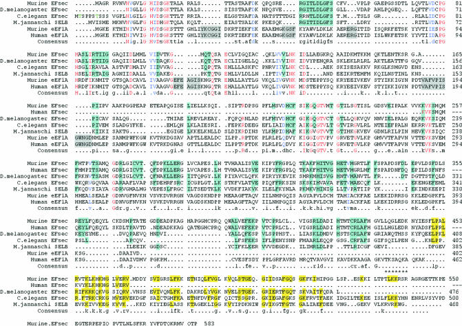
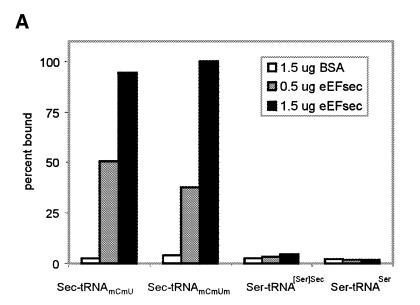
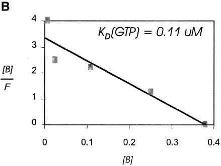
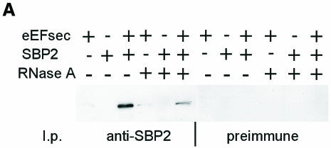
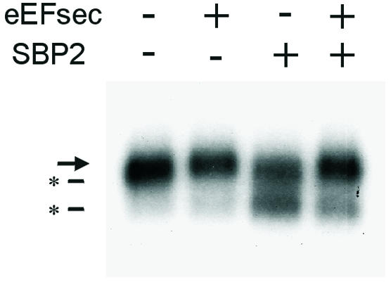
Decoding UGA as selenocysteine requires a unique tRNA, a specialized elongation factor, and specific secondary structures in the mRNA, termed SECIS elements. Eukaryotic SECIS elements are found in the 3' untranslated region of selenoprotein mRNAs while those in prokaryotes occur immediately downstream of UGA. Consequently, a single eukaryotic SECIS element can serve multiple UGA codons, whereas prokaryotic SECIS elements only function for the adjacent UGA, suggesting distinct mechanisms for recoding in the two kingdoms. We have identified and characterized the first eukaryotic selenocysteyl-tRNA-specific elongation factor. This factor forms a complex with mammalian SECIS binding protein 2, and these two components function together in selenocysteine incorporation in mammalian cells. Expression of the two functional domains of the bacterial elongation factor-SECIS binding protein as two separate proteins in eukaryotes suggests a mechanism for rapid exchange of charged for uncharged selenocysteyl-tRNA-elongation factor complex, allowing a single SECIS element to serve multiple UGA codons.


Figures

None
Fig. 1. Alignment of deduced amino acid sequences of eEF and eEF1A family members. Alignment was generated using Multalign software (Corpet, 1988). Red lettering indicates conserved amino acids. Blue lettering indicates similar amino acids. Light blue highlighting indicates regions conserved amongst the eEFsec family but differing from eEF1A. Gray highlighting indicates insertions in eEF1A relative to EFsec. Yellow highlighting indicates conserved or similar regions in the C-terminal eEFsec extensions. Asterisks indicate the predicted nuclear localization signal. Homology between murine eEFsec and D. melanogaster eEFsec is 41%, C.elegans is 30% and M. jannaschii is 33%.
None
Fig. 2. tRNA and guanine nucleotide binding specificity of murine eEFsec. (A) Binding of radiolabeled aminoacyl-tRNAs by the indicated proteins is given as percent total input counts retained on nitrocellulose filters. Seryl-tRNA[Ser]Sec was tested as either the individual mcmU and mcmUm isoacceptors or as a mixture of the two, with the same results. (B) Scatchard analysis of GTP binding to eEFsec. Binding was performed as described in Methods.
None
Fig. 3. Co-immunoprecipitation of murine eEFsec and SBP2 from co-transfected cells. (A) Co-immunoprecipitation of eEFsec with antisera against SBP2, followed by western blotting and detection with anti-FLAG (eEFsec) antibody. (B) Co-immunoprecipitation of truncated eEFsec-t as above. (B) was exposed ∼8-fold longer than (A). (C) Western analysis of eEFsec or eEFsec-t in total cell homogenates.
None
Fig. 4. Complex formation between eEFsec, SBP2 and SECIS element. (A) EMSA of w.t. type 1 deiodinase SECIS element (41 nucleotides of minimal SECIS element plus 35 nucleotides of flanking sequence). Arrows indicate shifted complexes, asterisks designate free RNA. (B) EMSA of indicated mutant SECIS elements corresponding to w.t. element above. (C) UV cross-linking of SBP2–SECIS and eEFsec–SBP2–SECIS complexes. The w.t. D1 SECIS element was incubated with the indicated proteins as for EMSA, followed by UV cross-linking, PAGE and western blotting as described in Methods. The arrow indicates the eEFsec–SBP2 complex.
None
Fig. 5. Effects of eEFsec and SBP2 on selenoprotein synthesis in vivo. Danio rerio selenoprotein P cDNA in pUHD10-3 vector was transfected into HEK-293 cells either alone or with plasmids expressing eEFsec, SBP2 or both as described in Methods. Twenty-four hours after transfection, [75Se]sodium selenite was added to the medium. Media were harvested the following day and analyzed by SDS–PAGE, followed by autoradiography. The arrow indicates the position of the full-length selenoprotein. Asterisks indicate truncated forms.

References

    1. Berry M.J., Banu, L., Chen, Y., Mandel, S.J., Kieffer, J.D., Harney, J.W. and Larsen, P.R. (1991a) Recognition of UGA as a selenocysteine codon in Type I deiodinase requires sequences in the 3′ untranslated region. Nature, 353, 273–276. - PubMed
    1. Berry M.J., Banu, L. and Larsen, P.R. (1991b) Type I iodothyronine deiodinase is a selenocysteine-containing enzyme. Nature, 349, 438–440. - PubMed
    1. Berry M.J., Banu, L., Harney, J.W. and Larsen, P.R. (1993) Functional characterization of the eukaryotic SECIS elements which direct selenocysteine insertion at UGA codons. EMBO J., 12, 3315–3322. - PMC - PubMed
    1. Böck A. (2000) Biosynthesis of selenoproteins—an overview. BioFactors, 11, 77–78. - PubMed
    1. Copeland P.R., Fletcher, J.E., Carlson, B.A., Hatfield, D.L. and Driscoll, D.M. (2000) A novel RNA binding protein, SBP2, is required for the translation of mammalian selenoprotein mRNAs. EMBO J., 19, 306–314. - PMC - PubMed

Publication types

MeSH terms