. 2001 Mar;2(3):222-8.
doi: 10.1093/embo-reports/kve046.

ASK1 is required for sustained activations of JNK/p38 MAP kinases and apoptosis

Affiliations

ASK1 is required for sustained activations of JNK/p38 MAP kinases and apoptosis

K Tobiume et al. EMBO Rep. 2001 Mar.

Abstract

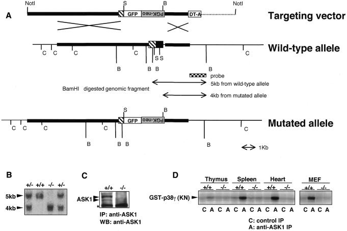
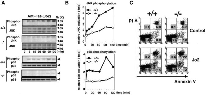
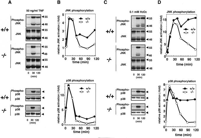
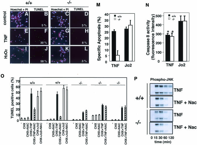
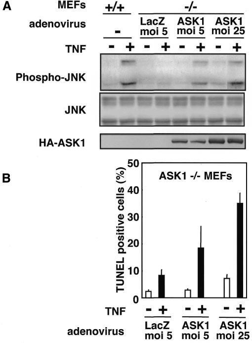
Apoptosis signal-regulating kinase (ASK) 1 is activated in response to various cytotoxic stresses including TNF, Fas and reactive oxygen species (ROS) such as H(2)O(2), and activates c-Jun NH(2)-terminal kinase (JNK) and p38. However, the roles of JNK and p38 signaling pathways during apoptosis have been controversial. Here we show that by deleting ASK1 in mice, TNF- and H(2)O(2)-induced sustained activations of JNK and p38 are lost in ASK1(-/-) embryonic fibroblasts, and that ASK1(-/-) cells are resistant to TNF- and H(2)O(2)-induced apoptosis. TNF- but not Fas-induced apoptosis requires ROS-dependent activation of ASK1-JNK/p38 pathways. Thus, ASK1 is selectively required for TNF- and oxidative stress-induced sustained activations of JNK/p38 and apoptosis.


Figures

None
Fig. 1. Generation of ASK1–/– mice by gene targeting. (A) Restriction map around the first coding exon of ASK1 gene and structure of the targeting vector. Homologous regions for recombination are drawn by thick lines in the restriction map. Coding and non-coding regions of ASK1 gene are represented by filled and hatched boxes, respectively. The genomic fragment used as a probe for Southern blot analysis for precise determination of recombination in ES cells and genotypes of mice is indicated below the map. The expected mutant allele and polymorphic fragments for genotyping from BamHI-digested genomic DNA are also indicated. GFP, coding sequence for green fluorescence protein followed by bovine growth hormone poly(A) signal; PGK-neo, coding sequence for neomycin phosphotransferase flanked by the phosphoglycerate kinase promoter and poly(A) signal; DT-A, diphtheria toxin A gene driven by MC1 promoter; C, SacI; S, SmaI; B, BamHI. Inverted characters for ‘PGK-neo’ indicate an insertion of the PGK-neo cassette in a reverse orientation. (B) Southern blot determining homologous recombination within the ASK1 locus of genomic DNA extracted from tails of F2 mice. BamHI digestion generated hybridized bands of the expected sizes for wild-type (5 kb) and mutant (4 kb). (C) Western blot demonstrating the absence of ASK1 protein in ASK1–/– MEFs. MEFs were immunoprecipitated with anti-ASK1 antiserum (DAV) and subsequently subjected to immunoblotting with anti-ASK1. Asterisk indicates non-specific reactions. (D) Immune complex kinase assay confirming the absence of ASK1 kinase activity in ASK1–/– mice. Extracts from indicated tissues and cells were immunoprecipitated with anti-ASK1 or control serum. Immune complex was incubated with GST–MKK6, and a kinase activity was then measured with the substrate GST–p38 γ(KN) which resulted in a phosphorylated image of GST–p38 γ (KN).
None
Fig. 2. Lack of JNK and p38 activation but not apoptotic alteration in ASK1–/– cells in response to anti-Fas (Jo2) treatment. (A) JNK and p38 activation in thymocytes. Cells were isolated from 6-week-old mice, preincubated in serum-free RPMI medium for 4 h and treated with 5 µg/ml Jo2 for the indicated period. Cell lysates were subjected to western blot analysis with antibodies specifically recognizing the activated forms of JNK (phospho-JNK) or p38 (phospho-p38). Membranes were reprobed with antibodies to total JNK or p38 as loading controls. (B) Graphic representation of (A). Fold activation was calculated by dividing the intensity of phosphorylated p55 JNK or p38 at different time points by that at time zero. Essentially similar results were obtained in three independent experiments. (C) Fluorescence analysis of apoptosis of Jo2-treated thymocytes from ASK1+/+ (left panels) or ASK1–/– mice (right panels). Cells were isolated from 6-week-old mice, suspended in serum-free RPMI medium, and cultured in the presence (lower panels) or absence (upper panels) of 5 µg/ml Jo2 for 8 h. Cells were analyzed by FACS after staining with Annexin V and propidium iodide (PI). Percentages of positive cells within a quadrant are indicated. Data are from one of more than five representative experiments.
None
Fig. 3. Lack of TNF- and H2O2-induced sustained activations of JNK and p38 in ASK1–/– MEFs. MEFs were treated with 50 ng/ml of TNF (A and B) or with 0.1 mM of H2O2 (C and D), and JNK and p38 activation was evaluated as described in Figure 2.
None
Fig. 4. Lack of TNF- and H2O2-induced apoptosis in ASK1–/– MEFs and differential requirement of ROS-ASK1 pathway in TNF- and Fas-induced apoptosis. Cell morphology (A, C, E, G, I, K) was analyzed by fluorescence double-staining with Hoechst 33258 (blue) and PI (red) of MEFs that were untreated (A and C), treated with 100 ng/ml of TNF for 8 h (E and G) or with 0.2 mM H2O2 plus 25 mM aminotriazole (catalase inhibitor) for 2h (I and K). TUNEL staining (green; B, D, F, H, J, L) of the same fields is shown with numbers which represent percentages of TUNEL-positive cells per total cell count. At least 100 cells from three random fields were counted in each experiment, and data are from one of more than three representative experiments. (M) Quantitative analysis of apoptosis of TNF- or Jo2-treated MEFs from ASK1+/+ or ASK1–/– mice. ASK1+/+ MEFs (black columns) and ASK1–/– MEFs (white columns) were treated with 100 ng/ml TNF plus 1 µg/ml cycloheximide (CHX), 5 µg/ml Jo2 plus 1 µg/ml CHX or 1 µg/ml CHX alone for 16 h. Apoptotic cells were scored by TUNEL staining as described above. Specific apoptosis was calculated as the percentage of TUNEL-positive cells in each experimental condition minus the percentage of TUNEL-positive cells with treatment with CHX alone. Data are from one of three representative experiments. (N) Quantitative analysis of caspase 8 activity in TNF- or Jo2-treated MEFs. ASK1+/+ MEFs (black columns) and ASK1–/– MEFs (white columns) were treated with 100 ng/ml TNF plus 1 µg/ml CHX or 5 µg/ml Jo2 plus 1 µg/ml CHX for 6 h. Caspase 8 activity was measured by a Caspase-8/FLICE Fluorometric Protease Assay Kit (MBL) using fluorogenic synthetic peptide IETD-7-amino-4-trifluoromethyl coumarin (AFC) as a substrate. The fluorescence intensity of the released AFC as caspase 8 activity was measured with an exitation wavelength of 360 nm and emission wavelength of 530 nm. Values are means (± SE) of four independent experiments. *, P <0.05 compared with Jo2-treated cells. (O) ASK1+/+ and ASK1–/– MEFs were treated with indicated combinations of CHX (1 µg/ml), Nac (1 mM), TNF (100 ng/ml) and Jo2 (5 µg/ml) for 24 h. TUNEL staining was performed, and the percentage of TUNEL-positive cells per total cell count in each experimental condition were calculated. Values are means (± SD) of three random fields (at least 100 cells per field counted). Results of MEFs prepared from independent embryos are shown. (P) Inhibition of TNF-induced sustained activation of JNK by Nac. ASK1+/+ and ASK1–/– MEFs were treated with 50 ng/ml of TNF in the presence or absence of Nac (1 mM), and cell lysates were subjected to western blot analysis with phospho-JNK antibody.
None
Fig. 5. Restoration of TNF sensitivity in ASK1-re-introduced ASK1–/– MEFs. (A) Restoration of TNF-induced sustained activation of JNK. ASK1–/– MEFs were infected with adenoviruses encoding β-galactosidase (LacZ) or HA-tagged wild-type ASK1 (Saitoh et al. 1998) for 24 h. Cells were treated with TNF (50 ng/ml for 120 min), and cell lysates were subjected to westen blot analysis with phospho-JNK antibody. Essentially the same relsults were obtained for H2O2-treatment. Appropriate expression of exogenous ASK1 is shown by anti-HA blotting. (B) Restoration of TNF-induced apoptosis. ASK1–/– MEFs were infected as described above, treated with TNF (50 ng/ml in the presence of 1 µg/ml CHX) for 6 h, and the percentage of TUNEL-positive cells was calculated as described in Figure 4O. Values are means (± SD) of three random fields (at least 100 cells per field counted).

References

    1. Chang H., Nishitoh, H., Yang, X., Ichijo, H. and Baltimore D. (1998) Activation of apoptosis signal-regulating kinase 1 (ASK1) by the death adaptor Daxx. Science, 281, 1860–1863. - PubMed
    1. Chen Y.-R., Wang, X., Templeton, D., Davis, R. and Tan, T.-H. (1996) The role of c-Jun N-terminal kinase (JNK) in apoptosis induced by ultraviolet C and γ-radiation. J. Biol. Chem., 50, 31929–31936. - PubMed
    1. Davis R.J. (2000) Signal transduction by the JNK group of MAP kinases. Cell, 103, 239–252. - PubMed
    1. Gross, A, McDonnell, J.M. and Korsmeyer, S.J. (1999) BCL-2 family members and the mitochondria in apoptosis. Genes Dev., 13, 1899–1911. - PubMed
    1. Guo Y.-L., Baysal, K., Kang, B., Yang, L.-J. and Williamson, J.R. (1998) Correlation between sustained c-Jun N-terminal protein kinase activation and apoptosis induced by tumor necrosis factor-α in rat mesangial cells. J. Biol. Chem., 273, 4027–4034. - PubMed

Publication types

MeSH terms

Substances