. 2001 Jan 22;152(2):349-60.
doi: 10.1083/jcb.152.2.349.

The Ndc80p complex from Saccharomyces cerevisiae contains conserved centromere components and has a function in chromosome segregation

Affiliations

The Ndc80p complex from Saccharomyces cerevisiae contains conserved centromere components and has a function in chromosome segregation

P A Wigge et al. J Cell Biol. .

Abstract

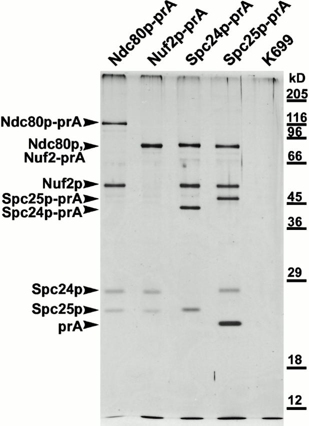
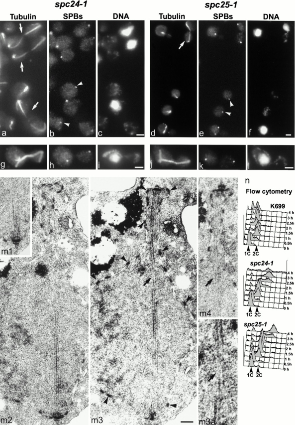
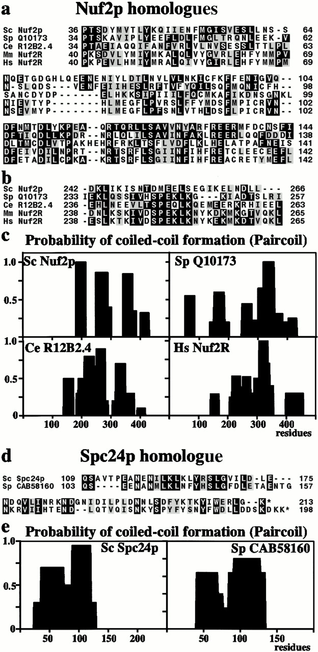
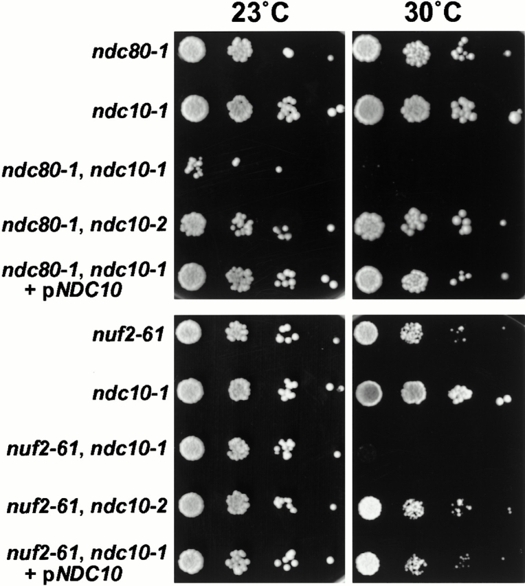
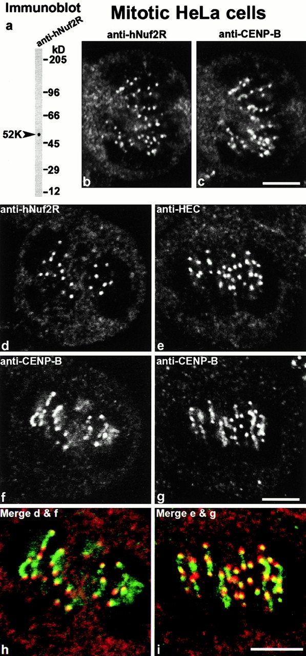
We have purified a complex from Saccharomyces cerevisiae containing the spindle components Ndc80p, Nuf2p, Spc25p, and Spc24p. Temperature-sensitive mutants in NDC80, SPC25, and SPC24 show defects in chromosome segregation. In spc24-1 cells, green fluorescence protein (GFP)-labeled centromeres fail to split during spindle elongation, and in addition some centromeres may detach from the spindle. Chromatin immunoprecipitation assays show an association of all four components of the complex with the yeast centromere. Homologues of Ndc80p, Nuf2p, and Spc24p were found in Schizosaccharomyces pombe and GFP tagging showed they were located at the centromere. A human homologue of Nuf2p was identified in the expressed sequence tag database. Immunofluorescent staining with anti-human Nuf2p and with anti-HEC, the human homologue of Ndc80p, showed that both proteins are at the centromeres of mitotic HeLa cells. Thus the Ndc80p complex contains centromere-associated components conserved between yeasts and vertebrates.


Figures

Figure 1
Figure 1
Silver-stained gel of the purified Ndc80p complex, prepared by tagging each of the individual components with prA and affinity purification on an IgG-Sepharose column. Components were identified by MALDI mass spectrometry (see Materials and Methods). Note that in the second lane, Ndc80p and Nuf2p-prA comigrate. The lane marked K699 shows an untagged strain.
Figure 4
Figure 4
Live cell imaging of spc24-1 cells containing GFP-Spc42p, an SPB component, and GFP-CEN5. CEN5 was labeled by integration of tet operators (which bind GFP-tet repressors) 1.4 kb from CEN5 (Tanaka et al. 2000). Cells were synchronized in G1 with α-factor and released at 23°C for 1.5 h (a) or at 36°C (b1–b3, c1–c2) for the times indicated; arrowheads show the SPBs which are brighter than the centromeres (He et al. 2000; Tanaka et al. 2000). The same cell is imaged in b1–b3 and another cell in c1–c2. Bar, 2 μm.
Figure 2
Figure 2
ImmunoEM of Nuf2p-GFP in short spindles (a and b) and a longer spindle in three consecutive serial sections (c1–c3). Bars, 0.1 μm.
Figure 3
Figure 3
Phenotype of spc24-1 and spc25-1. Immunofluorescent staining of unsynchronized spc24-1 (a–c) and spc25-1 cells (d–f) after 4 h at 36°C with antitubulin (a and d), anti-Tub4p (b and e), and DAPI (c and f). Arrowheads show aploid cells with SPBs, arrows postanaphase cells. spc24-1 (g–i) and spc25-1 (j–l) cells synchronized in G1 with α-factor and released at 36°C for 1.5 h were stained similarly. (m) EM of the same synchronized spc24-1 cells at 1.5 h. Four consecutive serial sections are shown (m1–m4), and overlapping microtubules are present between the two SPBs. The same SPB is shown in m1 and m2. Arrowheads show some of the nuclear pore complexes in m3, and the arrow shows an apparent discontinuity in the spindle in m3 caused by the microtubules being slightly out of the plane of the section. The microtubules can be seen in the equivalent position in the next section (arrow in m4) and even followed if viewed end on at twice the magnification of m3 (m3a). (n) Flow cytometry of K699 (wt), spc24-1, and spc25-1 cells synchronized in G1 and released at 36°C. Bars: (a–l) 2 μm; (m 1–4) 0.2 μm.
Figure 7
Figure 7
(a) Homologues of S. cerevisiae (Sc) Nuf2p in S. pombe (Sp), C. elegans (Ce), mice (Mm), and humans (Hs), showing homology in the mainly noncoiled coil NH2-terminal region. (b) In four of the homologues there is a break in the coiled coil at around residue 250 associated with an SPEKLK motif. (c) There is a broadly similar distribution of coiled coil domains in the COOH-terminal region of all four homologues. (d) Homologues of Spc24p in S. cerevisiae and S. pombe showing homology in the COOH-terminal region and a similar coiled coil distribution (e).
Figure 5
Figure 5
Synthetic growth defect in the ndc80-1, ndc10-1 and nuf2-61, ndc10-1 double mutants when cells were spotted on plates with increasing dilutions at 30°C. This effect was not observed when the ndc10-2 allele was used or the double mutants were transformed with a plasmid containing NDC10.
Figure 6
Figure 6
ChIP assay of strains containing prA-tagged Ndc80p complex components together with controls, prA-tagged Spc110p and Spc34p, and wt cells (K699). Regions of DNA amplified were across CEN3, across 1 kb to either side of CEN3, and across an AT-rich region (see Materials and Methods).
Figure 9
Figure 9
(a) Immunoblot of HeLa cell extract with anti-hNuf2R. Double label immunofluorescent staining by confocal microscopy of mitotic HeLa cells with anti-hNuf2R (b and d), anti-HEC (e), and anti–CENP-B (c, f, and g). Bars, 5 μm.
Figure 8
Figure 8
(a) Live cell imaging of SpNdc80p-GFP (b1 and b2), SpNuf2p-GFP (c1 and c2), and SpSpc24p-GFP (d1 and d2) in S. pombe. Cells about to complete anaphase A (b1, c1, and d1) show between five and six spots which within a few minutes coalesce into two spots during anaphase B (b2, c2, and d2). (e–g) Fluorescence of GFP compared with immunofluorescence with anti-Sad1 (e), antitubulin (f), and anti–HA-tagged Bub1 (g). Bars, 2.5 μm.

References

    1. Adams I.R., Kilmartin J.V. Localization of core spindle pole body (SPB) components during SPB duplication in Saccharomyces cerevisiae . J. Cell Biol. 1999;145:809–823. - PMC - PubMed
    1. Adams R.R., Wheatley S.P., Gouldsworthy A.M., Kandels-Lewis S.E., Carmena M., Smythe C., Gerloff D.L., Earnshaw W.C. INCENP binds the aurora-related kinase AIRK2 and is required to target it to chromosomes, the central spindle and cleavage furrow. Curr. Biol. 2000;10:1075–1078. - PubMed
    1. Aitchison J.D., Rout M.P., Marelli M., Blobel G., Wozniak R.W. Two novel related yeast nucleoporins Nup170p and Nup155pcomplementation with the vertebrate homologue Nup155p and functional interactions with the yeast nuclear pore-membrane protein Pom152p. J. Cell Biol. 1995;131:1133–1148. - PMC - PubMed
    1. Altschul S.F., Gish W., Miller W., Myers E.W., Lipman D.J. Basic local alignment search tool. J. Mol. Biol. 1990;215:403–410. - PubMed
    1. Bahler J., Wu J.Q., Longtine M.S., Shah N.G., McKenzie A., III, Steever A.B., Wach A., Philippsen P., Pringle J.R. Heterologous modules for efficient and versatile PCR-based gene targeting in Schizosaccharomyces pombe . Yeast. 1998;14:943–951. - PubMed

MeSH terms