. 2001 Jan 22;152(2):401-10.
doi: 10.1083/jcb.152.2.401.

Mmm1p, a mitochondrial outer membrane protein, is connected to mitochondrial DNA (mtDNA) nucleoids and required for mtDNA stability

Affiliations

Mmm1p, a mitochondrial outer membrane protein, is connected to mitochondrial DNA (mtDNA) nucleoids and required for mtDNA stability

A E Hobbs et al. J Cell Biol. .

Abstract

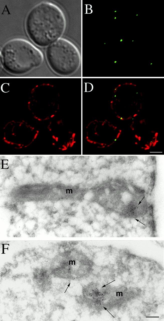
In the yeast Saccharomyces cerevisiae, mitochondria form a branched, tubular reticulum in the periphery of the cell. Mmm1p is required to maintain normal mitochondrial shape and in mmm1 mutants mitochondria form large, spherical organelles. To further explore Mmm1p function, we examined the localization of a Mmm1p-green fluorescent protein (GFP) fusion in living cells. We found that Mmm1p-GFP is located in small, punctate structures on the mitochondrial outer membrane, adjacent to a subset of matrix-localized mitochondrial DNA nucleoids. We also found that the temperature-sensitive mmm1-1 mutant was defective in transmission of mitochondrial DNA to daughter cells immediately after the shift to restrictive temperature. Normal mitochondrial nucleoid structure also collapsed at the nonpermissive temperature with similar kinetics. Moreover, we found that mitochondrial inner membrane structure is dramatically disorganized in mmm1 disruption strains. We propose that Mmm1p is part of a connection between the mitochondrial outer and inner membranes, anchoring mitochondrial DNA nucleoids in the matrix.


Figures

Figure 1
Figure 1
Mmm1p is localized in punctate structures on the mitochondrial surface. mmm1::URA3 strain YAAH1, which expresses Mmm1p-GFP from plasmid pAA2, was grown in YEP glycerol/ethanol media to early log phase and stained with MitoTracker™ Red. Live cells were mounted on slides and examined using a DeltaVision microscope system. 15 total images were taken in the z axis through the cells, deconvolved, and 7 of the 15 optical sections near the top of the cells were flattened into a single image. (A) Differential interference contrast (DIC); (B) Mmm1p-GFP fluorescence; (C) MitoTracker fluorescence; (D) merged images of B and C. Bar: 2 μm. (E and F) Strain YAAH3, which expresses Mmm1p-HA, was fixed, embedded, and frozen. Cryosections were incubated with antibodies to the HA epitope, followed by incubation with secondary antibodies conjugated to 5-nm gold particles. After staining, sections were examined under the electron microscope. Bar: 0.1 μm.
Figure 2
Figure 2
Mmm1p is adjacent to mitochondrial DNA nucleoids. Mmm1p-GFP–containing strain YAAH1 was grown to early log phase, stained with DAPI, and examined using a DeltaVision microscope. DAPI staining of live cells preferentially stains mtDNA, and the nucleus is only poorly stained (Williamson and Fennell 1979). Shown are all 15 deconvolved z sections, which have been flattened to a single image. (A) DIC; (B) Mmm1p-GFP fluorescence; (C) DAPI fluorescence; (D) merged images of B and C. (E–H) The images in D were processed using the DeltaVision model building tool, which removes background and shows fluorescent signals as solid objects for easier viewing. Four views of the 3-D model, each rotated 0° (E), 65° (F), 130° (G), and 190° (H) with respect to each other. Bar: 2 μm.
Figure 3
Figure 3
Mmm1p maintains a punctate localization in the absence of actin or mtDNA. (A) YAAH1, which expresses Mmm1p-GFP, was grown to early log phase and treated with Latrunculin A (+LatA), or mock treated (−LatA) at 24°C for 30 min. Cells were fixed and stained with Rhodamine-Phalloidin to visualize the actin cytoskeleton. Representative images of cells examined by fluorescence microscopy in the green (GFP) and red (Rh.-Phalloidin) channels are shown. (B) Mmm1p-GFP–expressing strains YAAH1, which contains mtDNA (ρ+) and YAAH2, which lacks mtDNA (ρ°), were stained with 1 μg/ml DAPI to visualize the mtDNA. Although DAPI treatment of living cells preferentially stains mtDNA, low-level staining of nuclear DNA can be seen when images are overexposed or no mtDNA is present. Bars: 2 μm.
Figure 4
Figure 4
mmm1-1 cells lose mtDNA at the nonpermissive temperature. (A) Wild-type strain RJ485 and mmm1-1 strain YSB105 were grown at 24°C in YEPD medium, and then shifted to 37°C for the indicated times. Total DNA was isolated, Southern blotted, and probed with 32P-labeled mitochondrial COX2 gene. After normalization to the levels of hybridization to the nuclear-encoded TIM23 gene, relative amounts of mtDNA in wild-type (□, solid line) and mmm1-1 (♦, dotted line) cells was plotted. (B) ade2 MMM1 strain RJ485 and ade2 mmm1-1 strain YSB105 were grown at 24°C in YEPD medium, and then shifted to 37°C for the indicated times and dilutions of cells were plated onto YEPD plates. After growth at 24°C for 3–5 d, the number of white (lacking functional mtDNA) and red (with functional mtDNA) colonies were determined. The percentage of white colonies using wild-type (□, solid line) and mmm1-1 (♦, dotted line) cells is indicated. (C) Wild-type and mmm1-1 cells were grown at 24°C, and then shifted to 37°C for the indicated times, stained with 1 μg/ml DAPI for 15 min, and examined by fluorescence microscopy. At each time point, 50 cells were examined and the percentage of cells with mtDNA is indicated. Wild-type (□, solid line) and mmm1-1 (♦, dotted line).
Figure 5
Figure 5
The number of mtDNA nucleoids rapidly decreases in mmm1-1 cells at the nonpermissive temperature. Wild-type strain RJ485 and mmm1-1 strain YSB105 were pregrown at 24°C, and then shifted to 37°C for the indicated times. After staining with DAPI, cells were examined by fluorescence microscopy. The number of individual mtDNA nucleoids per cell is plotted.
Figure 6
Figure 6
mtDNA nucleoids aggregate in mmm1-1 cells at the restrictive temperature. Wild-type and mmm1-1 cells were grown at 24°C, shifted to 37°C for the indicated times, and stained with DAPI. Cells were examined by fluorescence microscopy and representative fluorescent (left) and DIC (right) images are shown.
Figure 7
Figure 7
Normal inner membrane cristae morphology is lost in mmm1 disruption mutants. Wild-type (RJ485), wild-type rho° (YHS92), and mmm1::URA3 (RJ483) cells were grown at 24°C to mid-log phase, fixed, and thin sections were examined by electron microscopy. (A) Wild-type cells; (B) wild-type rhoo cells; (C) mmm1::URA3 cells. m, mitochondria; n, nucleus. Bar: 0.1 μm.
Figure 8
Figure 8
Mitochondrial membrane vesicles containing Mmm1p are located in a fraction intermediate in density to outer and inner membrane vesicles. Mitochondria were isolated from YSB107 (Burgess et al. 1994), which expresses the Mmm1p-HA fusion protein. Mitochondria were sonicated and membrane vesicles were separated on sucrose gradients (Pon et al. 1989). Gradient fractions were collected and analyzed by Western blotting using antibodies to Om45p, an outer membrane protein, the β-subunit of the F1-ATPase (F1β), an inner membrane protein, and HA (Mmm1p-HA). Lane 1 represents the top of the gradient.

References

    1. Adams A., Gottschling D.E., Kaiser C.A., Stearns T. Methods in Yeast Genetics 1997. Cold Spring Harbor Laboratories; Cold Spring Harbor, N.Y: pp. 1–112
    1. Ayscough K.R., Stryker J., Pokala N., Sanders M., Crews P., Drubin D.G. High rates of actin filament turnover in budding yeast and roles for actin in establishment and maintenance of cell polarity revealed using the actin inhibitor latrunculin-A. J. Cell Biol. 1997;137:399–416. - PMC - PubMed
    1. Azpiroz R., Butow R.A. Patterns of mitochondrial sorting in yeast zygotes. Mol. Biol. Cell. 1993;4:21–36. - PMC - PubMed
    1. Bereiter-Hahn J. Behavior of mitochondria in the living cell. Int. Rev. Cytol. 1990;122:1–62. - PubMed
    1. Bereiter-Hahn J., Voth M. Dynamics of mitochondria in living cellsshape changes, dislocations, fusion, and fission of mitochondria. Microsc. Res. Tech. 1994;27:198–219. - PubMed

Publication types

MeSH terms