. 2001 Feb 19;152(4):657-68.
doi: 10.1083/jcb.152.4.657.

Dissection of autophagosome formation using Apg5-deficient mouse embryonic stem cells

Affiliations

Dissection of autophagosome formation using Apg5-deficient mouse embryonic stem cells

N Mizushima et al. J Cell Biol. .

Abstract

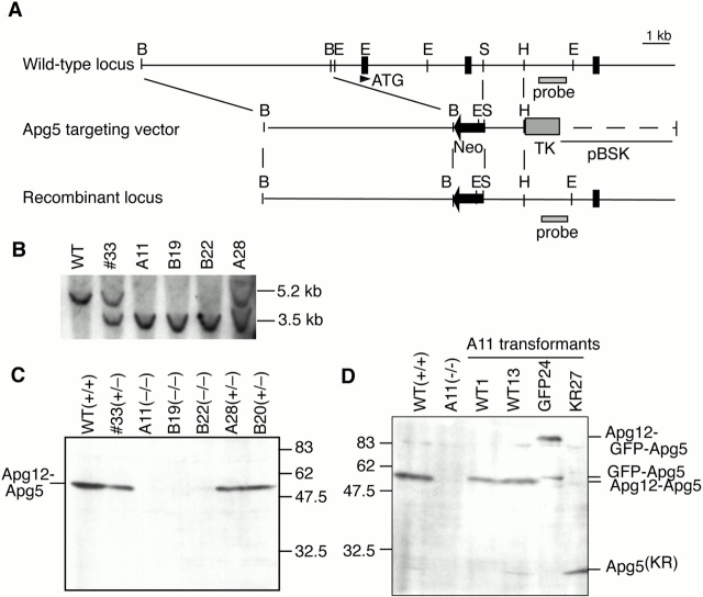
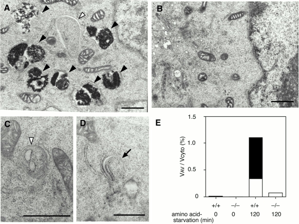
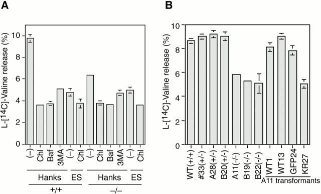
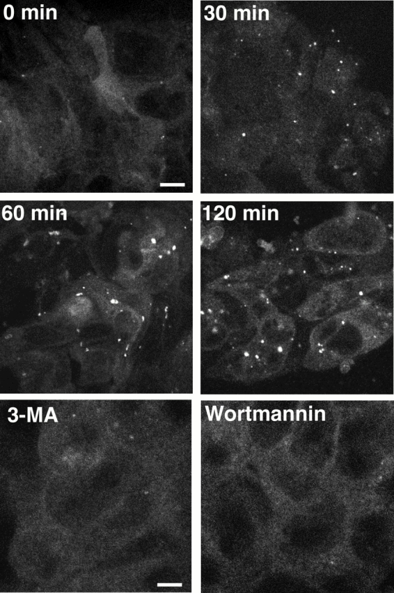
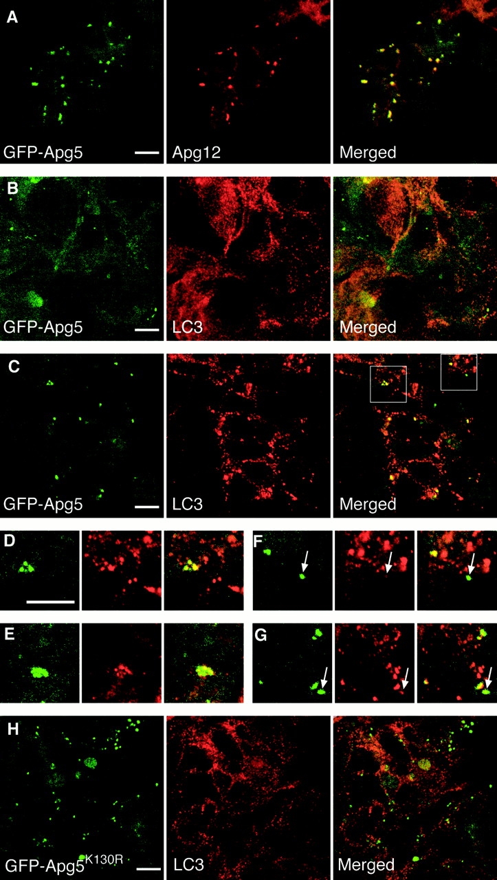
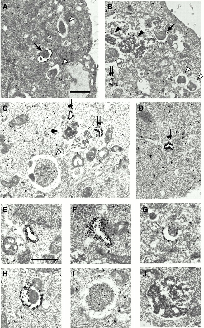
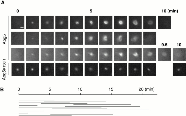
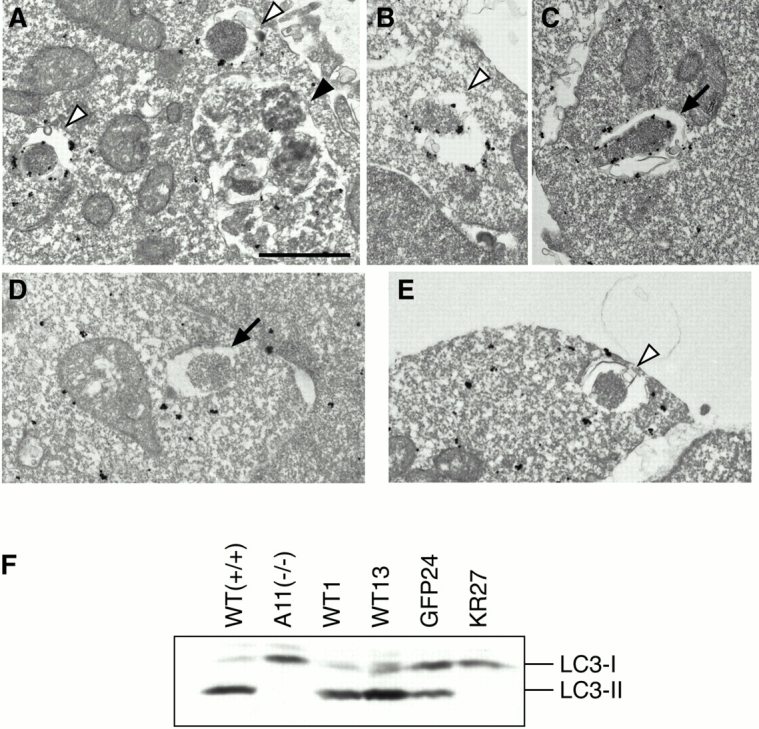
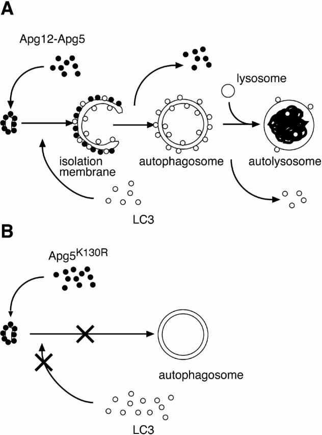
In macroautophagy, cytoplasmic components are delivered to lysosomes for degradation via autophagosomes that are formed by closure of cup-shaped isolation membranes. However, how the isolation membranes are formed is poorly understood. We recently found in yeast that a novel ubiquitin-like system, the Apg12-Apg5 conjugation system, is essential for autophagy. Here we show that mouse Apg12-Apg5 conjugate localizes to the isolation membranes in mouse embryonic stem cells. Using green fluorescent protein-tagged Apg5, we revealed that the cup-shaped isolation membrane is developed from a small crescent-shaped compartment. Apg5 localizes on the isolation membrane throughout its elongation process. To examine the role of Apg5, we generated Apg5-deficient embryonic stem cells, which showed defects in autophagosome formation. The covalent modification of Apg5 with Apg12 is not required for its membrane targeting, but is essential for involvement of Apg5 in elongation of the isolation membranes. We also show that Apg12-Apg5 is required for targeting of a mammalian Aut7/Apg8 homologue, LC3, to the isolation membranes. These results suggest that the Apg12-Apg5 conjugate plays essential roles in isolation membrane development.


Figures

Figure 1
Figure 1
Production of Apg5-deficient ES cells. (A) The restriction map of the wild-type APG5 allele, targeting construct, and mutated allele. Closed boxes indicate exons. Restriction enzymes: B, BamHI; E, EcoRI; S, SpeI; H, HindIII. (B) Southern blot analysis of wild-type ES cells (WT), an Apg5 single knockout clone (#33), three double knockout clones (A11, B19, and B22) and one single knockout clone obtained in the double knockout screening (A28). The probe indicated in A was used. (C) Immunoblot analysis of the ES clones. Total cell lysates were subjected to immunoblotting with anti–Apg5 antibody. Genotype of each clone is indicated. (D) Immunoblot analysis of stable transformants derived from the A11 clone with anti–Apg5 antibody.
Figure 2
Figure 2
APG5/− cells exhibit a block in the autophagic pathway. Wild-type ES cells (A) and APG5/− cells (B–D) were cultured in Hanks' solution for 2 h, and then fixed and subjected to conventional electron microscopic analysis. The isolation membrane (arrow), autophagosomes or autophagosome-like structures (open arrowheads), and autolysosomes (closed arrowheads) are indicated. (C and D) Rarely detected autophagosome-like structures in APG5/− cells. As often observed in wild-type cells, isolation membranes develop between cisternae of the ER (C and D). Bars, 1 μm. (E) Morphometric analysis of wild-type (+/+) and APG5/− (−/−) ES cells before or after amino acid withdrawal. Ratio of total area of autophagosomes (open column) and autolysosomes (closed column) to the total cytoplasmic area is shown.
Figure 3
Figure 3
Starvation-induced lysosomal protein degradation is reduced in APG5/−cells. (A) APG5 +/+ and APG5/− ES cells were labeled with l-[14C] valine for 24 h, and degradation of long-lived proteins during a 2-h incubation in Hanks' solution or the complete ES medium was measured as described in Materials and Methods. Chloroquine (Chl), bafilomycin A1 (Baf), or 3-MA was added as indicated. Data are the mean ± SD of triplicates from representative experiments. (B) ES clones and transformants were labeled with l-[14C] valine for 24 h and degradation of long-lived proteins during a 2-h incubation in Hanks' solution was measured.
Figure 4
Figure 4
Punctate signals of GFP-Apg5 increase under starvation conditions. (A) GFP24 cells were cultured in Hanks' solution for the times indicated or in Hanks' solution with 10 mM 3-MA or 100 nM wortmannin for 120 min and examined by confocal microscopy. Bars, 10 μm.
Figure 5
Figure 5
Apg12-Apg5 colocalizes with part of LC3. GFP24 (A–G) or GKR1 (H) cells were cultured in complete ES medium (B) or in Hanks' solution for 2 h (A and C–H). The cells were fixed, permeabilized, and subjected to immunofluorescence confocal microscopy using anti–mouse Apg12 antibody (A) or anti–LC3 antibody (B–H) and Cy5-conjugated goat anti–rabbit IgG antibody. GFP-Apg5 labeling (left), Apg12 or LC3 staining (middle), and merged images (right) are shown. (D and F) Higher magnifications of images shown in C. E and G are from another image. (F and G) Arrows indicate structures showing high levels of GFP-Apg5 but low levels of LC3. Bars, 10 μm.
Figure 6
Figure 6
GFP-Apg5 is present on isolation membranes. GFP24 (A–C, E–J) or GKR-1 (D) cells were cultured in Hanks' solution for 2 h and fixed. The localization of GFP-Apg5 was examined by silver-enhanced immunogold electron microscopy using an anti–GFP antibody. In B, an isolation membrane is enclosing a mitochondrion. The isolation membranes (arrows), autophagosomes (open arrowheads) and autolysosomes (closed arrowheads) are indicated. Double arrows indicate small membrane compartments to which GFP-Apg5 extensively localizes. The typical images of these structures are shown at higher magnification (E–J). Bars, 1 μm.
Figure 7
Figure 7
Formation of autophagosomes is traced with GFP-Apg5. (A) Sequential frames (1-min intervals) of three newly generated GFP-Apg5 spots, or a GFP-Apg5K130R spot. GFP24 or GKR-1 cells were cultured in Hanks' solution for 1 h and directly observed by time-lapse video microscopy. Bars, 1 μm. See supplemental videos of GFP-Apg5 (videos 1–3) and GFP-Apg5K130R (video 4). (B) Duration of each spot of GFP-Apg5 staining was measured for 15 cases that showed circularization in the same field.
Figure 8
Figure 8
LC3 does not target to membrane in APG5/− cells. Wild-type ES cells (A–C) and APG5/− cells (D and E) were cultured in Hanks' solution for 2 h and fixed. Localization of LC3 was examined by silver-enhanced immunogold electron microscopy using an antibody against recombinant LC3. The isolation membrane (arrow), autophagosomes (open arrowheads) and autolysosome (closed arrowhead) are indicated. Bar, 1 μm. (F) Total lysates from APG5 +/+ and APG5 −/− cells and their various stable transformants cultured in Hanks' solution for 2 h were subjected to immunoblot analysis with anti–LC3 antibody. Positions of LC3-I and LC3-II are indicated.
Figure 9
Figure 9
Model of subcellular localization of Apg12-Apg5 and LC3, and role of Apg12 conjugation in autophagosome formation. (A) Apg12-Apg5 conjugate localizes to the crescent-shaped autophagosome precursors. While these structures elongate and maturate into cup-shaped isolation membranes, LC3 is recruited to the membrane in the Apg5-dependent manner and Apg5 changes its localization to the outer side of the membrane. Apg5 plays an essential role in this membrane development. Immediately before or after the completion of autophagosome formation, Apg5 detaches from the membrane. Some LC3 also dissociate from the autophagosomal membrane thereafter. (B) Apg12 conjugation is not required for membrane targeting of Apg5, but it is essential for maturation of the isolation membrane into autophagosome and recruitment of LC3 to the membrane.

Similar articles

Cited by

References

    1. Baba M., Ohusumi M., Ohsumi Y. Analysis of the membrane structure involved in autophagy in yeast by freeze-replica method. Cell Struct. Funct. 1995;20:465–471. - PubMed
    1. Bolender R.P., Weibel E.R. A morphometric study of the removal of phenobarbital-induced membranes from hepatocytes after cessation of treatment. J. Cell Biol. 1973;56:746–761. - PMC - PubMed
    1. Clarke P.G. Developmental cell deathmorphological diversity and multiple mechanisms. Anat. Embryol. 1990;181:195–213. - PubMed
    1. Dice J.F. Peptide sequences that target cytosolic proteins for lysosomal proteolysis. Trends Biol. Sci. 1990;15:305–309. - PubMed
    1. Dunn W.A. Studies on the mechanisms of autophagyformation of the autophagic vacuole. J. Cell Biol. 1990;110:1923–1933. - PMC - PubMed

Publication types