. 2001 Feb 19;152(4):669-82.
doi: 10.1083/jcb.152.4.669.

Drosophila aurora B kinase is required for histone H3 phosphorylation and condensin recruitment during chromosome condensation and to organize the central spindle during cytokinesis

Affiliations

Drosophila aurora B kinase is required for histone H3 phosphorylation and condensin recruitment during chromosome condensation and to organize the central spindle during cytokinesis

R Giet et al. J Cell Biol. .

Abstract

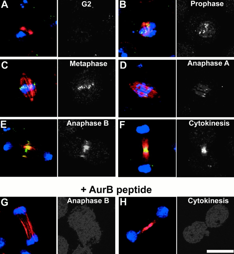
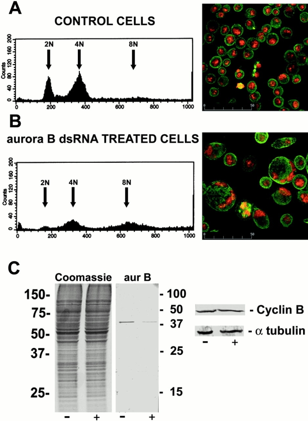
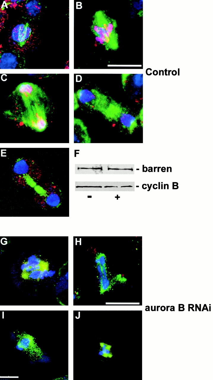
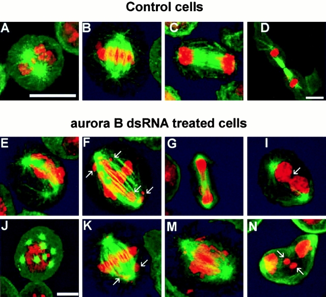
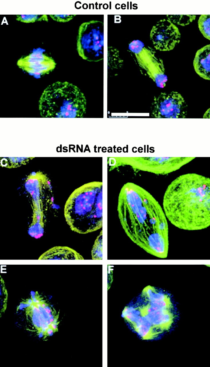
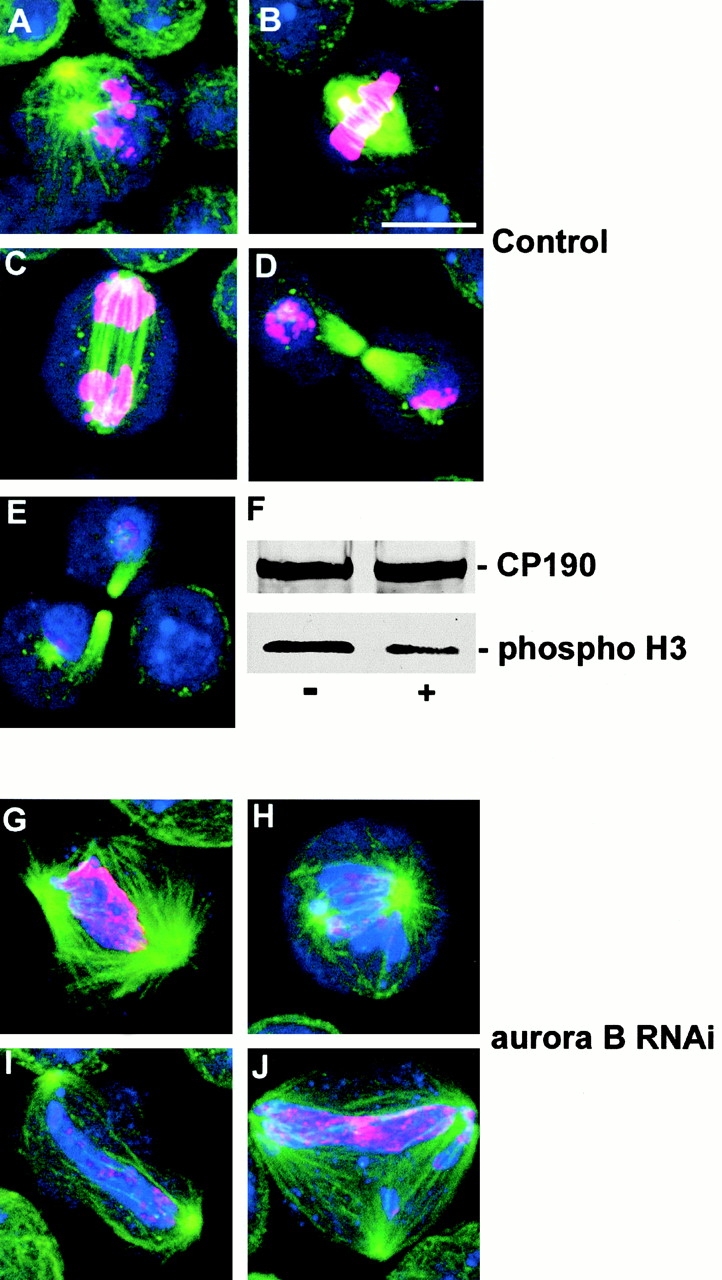
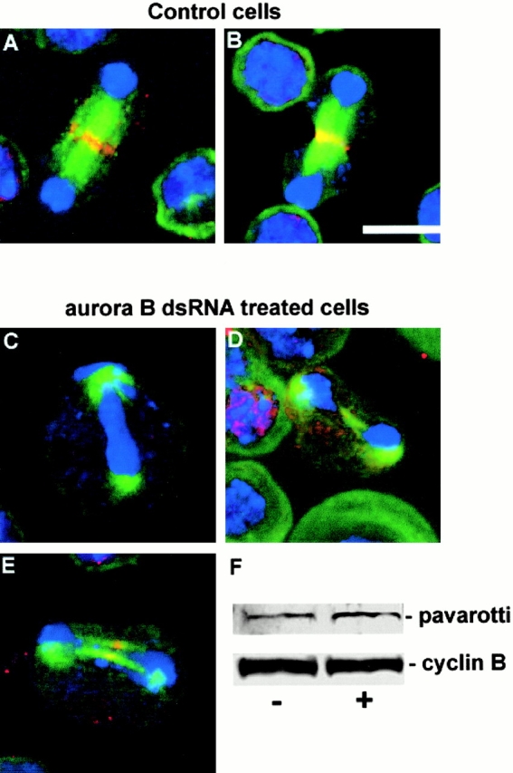
Aurora/Ipl1-related kinases are a conserved family of enzymes that have multiple functions during mitotic progression. Although it has been possible to use conventional genetic analysis to dissect the function of aurora, the founding family member in Drosophila (Glover, D.M., M.H. Leibowitz, D.A. McLean, and H. Parry. 1995. Cell. 81:95-105), the lack of mutations in a second aurora-like kinase gene, aurora B, precluded this approach. We now show that depleting Aurora B kinase using double-stranded RNA interference in cultured Drosophila cells results in polyploidy. aurora B encodes a passenger protein that associates first with condensing chromatin, concentrates at centromeres, and then relocates onto the central spindle at anaphase. Cells depleted of the Aurora B kinase show only partial chromosome condensation at mitosis. This is associated with a reduction in levels of the serine 10 phosphorylated form of histone H3 and a failure to recruit the Barren condensin protein onto chromosomes. These defects are associated with abnormal segregation resulting from lagging chromatids and extensive chromatin bridging at anaphase, similar to the phenotype of barren mutants (Bhat, M.A., A.V. Philp, D.M. Glover, and H.J. Bellen. 1996. Cell. 87:1103-1114.). The majority of treated cells also fail to undertake cytokinesis and show a reduced density of microtubules in the central region of the spindle. This is accompanied by a failure to correctly localize the Pavarotti kinesin-like protein, essential for this process. We discuss these conserved functions of Aurora B kinase in chromosome transmission and cytokinesis.


Figures

Figure 1
Figure 1
Aurora B is a chromosome passenger protein. Cultured Drosophila S2 cells were grown on glass coverslips, fixed, and stained to reveal DNA (blue), microtubules (red), and Aurora B kinase (green) (see Materials and Methods). Images are shown in pairs with the merged image on the left and the channel showing anti–Aurora B staining alone in monochrome on the right. The indicated successive stages of mitotic progression are shown in A–F. A detailed description of the Aurora B localization pattern is given in the text. In brief, it is first found throughout the condensing chromatin, concentrates at centromeres, and is then redistributed to the central spindle and midbody in late mitosis. G and H show cells stained in the presence of the peptide used to raise the anti–Aurora B antibody. Bar, 10 μm.
Figure 2
Figure 2
Polyploidy in Drosophila S2 cells treated with aurB dsRNA. FACS® profiles (left) and immunostaining (right) of control (A) or aurB RNAi (B) in S2 cells 3 d after addition of dsRNA to the experimental culture. Cells were stained to reveal DNA (red) and microtubules (green). (C) Protein profiles of similar preparations of S2 cells. Total Coomassie-stained proteins separated by SDS-PAGE are shown on the left, together with Western blots revealing Aurora B, cyclin B, and α-tubulin as indicated.
Figure 6
Figure 6
Barren fails to localize to mitotic chromosomes after depletion of Aurora B. (A–E) The cycle of association of Barren protein (red) onto mitotic chromosomes in control S2 cells. DNA is stained blue and microtubules green. Cells are shown at prophase (A), metaphase (B), anaphase (C), telophase (D), and cytokinesis (E). Association of Barren with chromosomes follows the same dynamics as histone H3 phosphorylation (see text). (F) Western blot showing that levels of Barren protein (top) are unchanged from control cells (−) after aurB RNAi (+) as are levels of cyclin B (bottom). (G–J) Abnormal mitotic figures in aurB RNAi cells in which barren does not localize to chromosomes. Bars, 10 μm.
Figure 3
Figure 3
Mitotic abnormalities in S2 cells depleted of Aurora B. (A–D) Control cells at prophase, metaphase, anaphase, and cytokinesis, respectively. Microtubules are stained green and DNA red. (E–N) Abnormal mitoses in aurora B–depleted cells in states resembling prophase (E and J), metaphase (F and K), anaphase (G and M), and telophase (I and N). The arrows in F and K indicate chromosomes that have not aligned on the metaphase plate. The arrows in I and N indicate a conspicuous chromatin bridge or lagging chromatids at telophase, respectively. Bars, 10 μm.
Figure 4
Figure 4
Centromere segregation is defective in Aurora B–depleted cells. (A and B) Control cells at metaphase and anaphase, respectively. (C–F) Abnormal mitoses in aurora B–depleted cells in an anaphase-like state showing centromeric regions (stained red with anti-Prod antibody) that fail to reach the spindle poles. DNA is stained blue and microtubules green. Bar, 10 μm.
Figure 5
Figure 5
Histone H3 phosphorylation in untreated and aurB RNAi S2 cells. (A–E) The cycle of histone H3 phosphorylation revealed by antiphospho-histone H3 antibody (red) in control S2 cells. DNA is stained blue and microtubules green. Cells are shown at prophase (A), metaphase (B), anaphase (C), and telophase/cytokinesis (D and E). Immunostaining of the phosphoepitope builds to a maximum during metaphase and early anaphase (see text). (F) A Western blot that shows reduction in phospho-histone H3 staining (bottom) in dsRNA-treated cells (+) compared with control cells (−), whereas CP190 levels (top) remain constant. (G–J) Abnormal mitotic figures in aurB RNAi cells showing reduced levels of phospho-histone H3 staining compared with control mitoses. Bar, 10 μm.
Figure 7
Figure 7
Failure of Pav-KLP to fully localize to the central spindle in Aurora B–depleted cells. (A and B) Pav-KLP (red) localizes to the central spindle/midbody in control S2 cells at late anaphase (A) and early cytokinesis (B). DNA is blue and microtubules are green. (C–E) Abnormal late mitotic figures in Aurora B–depleted cells in which the density of microtubules in the central spindle region is greatly reduced and in which the Pav-KLP either fails to localize (C) or localizes very poorly (D and E). (F) Western blot showing that levels of Pav-KLP (top) are unchanged from control cells (−) after treatment with aurB dsRNA (+), as are levels of cyclin B (bottom). Bar, 10 μm.

References

    1. Adams R.R., Tavers A.A., Salzberg A., Bellen H.J., Glover D.M. pavarotti encodes a kinesin-like protein required to organize the central spindle and contractile ring for cytokinesis. Genes Dev. 1998;12:1483–1494. - PMC - PubMed
    1. Adams R.R., Wheatleya S.P., Gouldsworthy A.M., Kandels-Lewis S.E., Carmena M., Smythe C., Gerloff D.L., Earnshaw W.C. INCENP binds the aurora-related kinase AIRK2 and is required to target it to chromosomes, the central spindle and cleavage furrow. Curr. Biol. 2000;10:1075–1078. - PubMed
    1. Bhat M.A., Philp A.V., Glover D.M., Bellen H.J. Chromatid segregation at anaphase requires the barren product, a novel chromosome-associated protein that interacts with Topoisomerase II. Cell. 1996;87:1103–1114. - PubMed
    1. Biggins S., Severin F.F., Bhalla N., Sassoon I., Hyman A.A., Murray A.W. The conserved protein kinase Ipl1 regulates microtubule binding to kinetochores in budding yeast. Genes Dev. 1999;13:532–544. - PMC - PubMed
    1. Bradbury E.M. Reversible histone modifications and the chromosome cell cycle. Bioessays. 1992;14:9–16. - PubMed

Publication types

MeSH terms