. 2001 Feb 19;152(4):717-28.
doi: 10.1083/jcb.152.4.717.

A specific role of phosphatidylinositol 3-kinase gamma. A regulation of autonomic Ca(2)+ oscillations in cardiac cells

Affiliations

A specific role of phosphatidylinositol 3-kinase gamma. A regulation of autonomic Ca(2)+ oscillations in cardiac cells

C Bony et al. J Cell Biol. .

Abstract

Purinergic stimulation of cardiomyocytes turns on a Src family tyrosine kinase-dependent pathway that stimulates PLCgamma and generates IP(3), a breakdown product of phosphatidylinositol 4,5-bisphosphate (PIP2). This signaling pathway closely regulates cardiac cell autonomic activity (i.e., spontaneous cell Ca(2+) spiking). PIP2 is phosphorylated on 3' by phosphoinositide 3-kinases (PI3Ks) that belong to a broad family of kinase isoforms. The product of PI3K, phosphatidylinositol 3,4,5-trisphosphate, regulates activity of PLCgamma. PI3Ks have emerged as crucial regulators of many cell functions including cell division, cell migration, cell secretion, and, via PLCgamma, Ca(2+) homeostasis. However, although PI3Kalpha and -beta have been shown to mediate specific cell functions in nonhematopoietic cells, such a role has not been found yet for PI3Kgamma. We report that neonatal rat cardiac cells in culture express PI3Kalpha, -beta, and -gamma. The purinergic agonist predominantly activates PI3Kgamma. Both wortmannin and LY294002 prevent tyrosine phosphorylation, and membrane translocation of PLCgamma as well as IP(3) generation in ATP-stimulated cells. Furthermore, an anti-PI3Kgamma, but not an anti-PI3Kbeta, injected in the cells prevents the effect of ATP on cell Ca(2+) spiking. A dominant negative mutant of PI3Kgamma transfected in the cells also exerts the same action. The effect of ATP was observed on spontaneous Ca(2+) spiking of wild-type but not of PI3Kgamma(2/2) embryonic stem cell-derived cardiomyocytes. ATP activates the Btk tyrosine kinase, Tec, and induces its association with PLCgamma. A dominant negative mutant of Tec blocks the purinergic effect on cell Ca(2+) spiking. Tec is translocated to the T-tubes upon ATP stimulation of cardiac cells. Both an anti-PI3Kgamma antibody and a dominant negative mutant of PI3Kgamma injected or transfected into cells prevent the latter event. We conclude that PI3Kgamma activation is a crucial step in the purinergic regulation of cardiac cell spontaneous Ca(2+) spiking. Our data further suggest that Tec works in concert with a Src family kinase and PI3Kgamma to fully activate PLCgamma in ATP-stimulated cardiac cells. This cluster of kinases provides the cardiomyocyte with a tight regulation of IP(3) generation and thus cardiac autonomic activity.


Figures
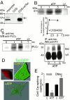
Figure 3
Figure 3
Tec is involved in ATP-induced slowing of cell Ca2+ spiking rate. (A) Whole cell lysate proteins from cardiac cells, mock Cos, or Cos cells transfected with wild-type Tec (from left to right lane), were subjected to Western blotting using an anti-Tec antibody. (B) After ATP stimulation, Tec was immunoprecipitated. The immunocomplex was used for the autophosphorylation assay using [γ-32P]ATP in the presence of MnCl2 and was analyzed by autoradiography. Cells were pretreated for 10 min with LY294002 before ATP stimulation (top). Data from three experiments are gathered in the line graph (middle). After Tec immunoprecipitation, a Western blot antiphosphotyrosine was performed to confirm the identity of the Tec amino acid phosphorylated after purinergic stimulation of cells (bottom). (C) After immunoprecipitation of Tec, the immunocomplex was subjected to Western blotting using an anti-PLCγ antibody to detect the presence of PLCγ. Similar experiments were repeated three times. (D) A TecGFP plasmid was transfected into cardiomyocytes. 36 h later, transfected cells were stimulated (ATP) or not (control) with 20 μM ATP. The inset shows the labeling of a cardiomyocyte by the anti-Tec antibody and a secondary TRITC-conjugated antibody. Specificity of the labeling was confirmed by a staining of cardiomyocytes overexpressing a wild-type Tec. The images were obtained after digital deconvolution (Huygens software) and visualized using a shadow projection (Imaris software). In this series of experiments 85 ± 5% of cells featured a membrane staining of TecGFP (E), mock cells or cells transfected with a dominant negative (DN) mutant of Tec were loaded with fluo3 and superfused with ATP. Fluo3 fluorescence was recorded in a region of interest including the whole cell with a CCD camera. Two to three images/s were captured by a CCD camera. A similar result was obtained in 12 cells isolated from two separate cultures as shown in the bar graph. *Significantly decreased; ^significantly increased (p ≤ 0.01). wt, Wild-type; ATP, ATP-stimulated cells; C, control.
Figure 1
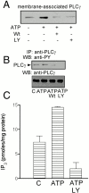
Figure 1
PI3K is required for ATP-induced IP3 generation. Cardiomyocytes were stimulated with 20 μM ATP in the absence or presence of wortmannin (Wt) or LY294002 (LY). After stimulation, cells were subfractionated in cytosolic and membrane fractions (A). Membrane proteins were subjected to Western blot analysis using an anti-PLCγ antibody. (B) PLCγ was immunoprecipitated, and the immunocomplex was analyzed by Western blot using an anti-phosphotyrosine or an anti-PLCγ antibody. (C) The amount of IP3 was assayed in a protein-free cell extract. The figure is representative of at least three experiments performed on three separate cell cultures.WB, Western blotting.
Figure 2
Figure 2
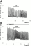
PI3K is required for ATP-induced slowing of cell Ca2+ spiking rate. A fluo3-loaded cell was superfused in the absence (A) or presence (B) of LY294002 with 20 μM ATP. Fluo3 fluorescence was recorded every 30 ms with a photomultiplier. The figure is representative of 10 similar experiments performed using two different cell cultures. Data are compared in the bar graph shown in (C). ^Significantly increased. (p ≤ 0.01). ATP, ATP-stimulated cells; C, control.
Figure 2
Figure 2
PI3K is required for ATP-induced slowing of cell Ca2+ spiking rate. A fluo3-loaded cell was superfused in the absence (A) or presence (B) of LY294002 with 20 μM ATP. Fluo3 fluorescence was recorded every 30 ms with a photomultiplier. The figure is representative of 10 similar experiments performed using two different cell cultures. Data are compared in the bar graph shown in (C). ^Significantly increased. (p ≤ 0.01). ATP, ATP-stimulated cells; C, control.
Figure 4
Figure 4
ATP specifically activates PI3Kγ. (A) PI3K subunits p85, p110α, -β and -γ were immunoprecipitated from a cardiac whole cell lysate and subjected to Western blot analysis using either an anti-p110 α1, β3, or γ antibody. The polyclonal anti-p110γ antibody was used for the immunoprecipitation and the monoclonal anti-p110γ antibody was used for Western blot. (B) Cells were stimulated for 0, 0.5, 1, or 5 min with 20 μM ATP, and PI3 kinase activity was assayed after PI3K immunoprecipitation. The experiment was repeated at least three times on three separate cultures. Means ± SEM of fold activation of PI3Ks are plotted as a function of the duration of purinergic stimulation of cells after scanning the autoradiographic films exposed to TLC plates (bottom).
Figure 6
Figure 6
Proposed signaling pathway of the purinergic receptor: regulation of cardiac autonomic activity. Activation of the G protein–coupled P2 purinergic receptor leads to dissociation of α and βγ subunits. α activates Fyn, whereas βγ stimulates PI3Kγ. Tec is then transphosphorylated by Fyn. This leads to phosphorylation and membrane translocation of PLCγ. PIP3, the product of PI3K, facilitates the anchor of both Tec and PLCγ. The latter is fully activated, generating IP3, which regulates the autonomic activity of the cardiomyocyte.
Figure 5
Figure 5
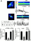
PI3Kγ mediates both ATP-induced slowing of cell Ca2+ spiking and Tec translocation to the cell membrane. (A) Cardiomyocytes were microinjected with a specific anti PI3Kβ or -γ polyclonal antibody together with fluo3. Cell Ca2+ spiking was monitored with a CCD camera. After background subtraction, a line was set offline along a cell (arrow). Fluorescence was recorded along the line as a function of time. The line scan images of adjacent lines were reconstructed using ANALYZE software (Mayo Foundation). The graphs below the images represent the change in fluorescence (ΔF) along the time after subtraction of the first image (Fo). Similar results were obtained on at least 10 cells from two separate cell cultures as shown in the bar graph on the left. Cells were also transfected with a dominant negative mutant (K399R) of PI3Kγ, and the effects of ATP on cell Ca2+ spiking was measured 24 h later on 10 different cells from two separate cultures (right). A recombinant p110γ was expressed as a GST-p110γ fusion (inset). PIP3K activity was then measured in vitro in the presence of rabbit IgG, the specific polyclonal anti-p110γ, or the anti-p110α or -p110β antibodies. (B) Beating embryoid bodies (days 9 and 10) generated from PI3Kγ2/2, PI3Kγ1/2, or wild-type (WT) ES cells were loaded with fluo3, and the effect of ATP was tested on spontaneous Ca2+ spiking of cardiomyocytes. The results are expressed as means ± SEM from 15 PI3Kγ2/2, 6 PI3Kγ1/2, and 6 wild-type embryoid bodies. (C) A specific anti-PI3Kβ (left) or γ antibody (middle) was injected, or a dominant negative mutant (K399R) of PI3Kγ was transfected into cells (right). Cardiomyocytes transfected with a TecGFP plasmid were microinjected. 1 h later, cells were stimulated for 1 min with ATP and fixed before image acquisition. Similar results were obtained in 15 cells from three separate cell cultures, as shown in the bar graph on left. Cells were also transfected with a dominant negative mutant (K399R) of PI3Kγ, and the effects of ATP on Tec membrane translocation was tested on eight transfected or mock cells (right). *Significantly decreased; ^significantly increased (p ≤ 0.01). ATP, ATP-stimulated cells; C, control.
Figure 5
Figure 5
PI3Kγ mediates both ATP-induced slowing of cell Ca2+ spiking and Tec translocation to the cell membrane. (A) Cardiomyocytes were microinjected with a specific anti PI3Kβ or -γ polyclonal antibody together with fluo3. Cell Ca2+ spiking was monitored with a CCD camera. After background subtraction, a line was set offline along a cell (arrow). Fluorescence was recorded along the line as a function of time. The line scan images of adjacent lines were reconstructed using ANALYZE software (Mayo Foundation). The graphs below the images represent the change in fluorescence (ΔF) along the time after subtraction of the first image (Fo). Similar results were obtained on at least 10 cells from two separate cell cultures as shown in the bar graph on the left. Cells were also transfected with a dominant negative mutant (K399R) of PI3Kγ, and the effects of ATP on cell Ca2+ spiking was measured 24 h later on 10 different cells from two separate cultures (right). A recombinant p110γ was expressed as a GST-p110γ fusion (inset). PIP3K activity was then measured in vitro in the presence of rabbit IgG, the specific polyclonal anti-p110γ, or the anti-p110α or -p110β antibodies. (B) Beating embryoid bodies (days 9 and 10) generated from PI3Kγ2/2, PI3Kγ1/2, or wild-type (WT) ES cells were loaded with fluo3, and the effect of ATP was tested on spontaneous Ca2+ spiking of cardiomyocytes. The results are expressed as means ± SEM from 15 PI3Kγ2/2, 6 PI3Kγ1/2, and 6 wild-type embryoid bodies. (C) A specific anti-PI3Kβ (left) or γ antibody (middle) was injected, or a dominant negative mutant (K399R) of PI3Kγ was transfected into cells (right). Cardiomyocytes transfected with a TecGFP plasmid were microinjected. 1 h later, cells were stimulated for 1 min with ATP and fixed before image acquisition. Similar results were obtained in 15 cells from three separate cell cultures, as shown in the bar graph on left. Cells were also transfected with a dominant negative mutant (K399R) of PI3Kγ, and the effects of ATP on Tec membrane translocation was tested on eight transfected or mock cells (right). *Significantly decreased; ^significantly increased (p ≤ 0.01). ATP, ATP-stimulated cells; C, control.

References

    1. August A., Sadra A., Dupont B., Hanafusa H. Src-induced activation of inducible T cell kinase (ITK) requires phosphatidylinositol 3-kinase activity and the Pleckstrin homology domain of inducible T cell kinase. Proc. Natl. Acad. Sci. USA. 1997;94:11227–11232. - PMC - PubMed
    1. Benistant C., Chapuis H., Roche S. A specific function for phosphatidylinositol 3-kinase α (p85-p110α) in cell survival and for phosphatidylinositol 3-kinase β (p85-p110β) in de novo DNA synthesis of human colon carcinoma cells. Oncogene. 2000;19:5083–5093. - PubMed
    1. Berridge M.J. Inositol trisphosphate and calcium signaling. Ann. NY Acad. Sci. 1995;766:31–43. - PubMed
    1. Clapham D.E. Calcium signaling. Cell. 1995;80:259–268. - PubMed
    1. Downing G.J., Kim S., Nakanishi S., Catt K.J., Balla T. Characterization of a soluble adrenal phosphatidylinositol 4-kinase reveals wortmannin sensitivity of type III phosphatidylinositol kinases. Biochemistry. 1996;35:3587–3594. - PubMed

Publication types

MeSH terms

Substances