. 2001 Feb 19;152(4):777-84.
doi: 10.1083/jcb.152.4.777.

The plasminogen activator inhibitor PAI-1 controls in vivo tumor vascularization by interaction with proteases, not vitronectin. Implications for antiangiogenic strategies

Affiliations

The plasminogen activator inhibitor PAI-1 controls in vivo tumor vascularization by interaction with proteases, not vitronectin. Implications for antiangiogenic strategies

K Bajou et al. J Cell Biol. .

Abstract

The plasminogen (Plg)/plasminogen activator (PA) system plays a key role in cancer progression, presumably via mediating extracellular matrix degradation and tumor cell migration. Consequently, urokinase-type PA (uPA)/plasmin antagonists are currently being developed for suppression of tumor growth and angiogenesis. Paradoxically, however, high levels of PA inhibitor 1 (PAI-1) are predictive of a poor prognosis for survival of patients with cancer. We demonstrated previously that PAI-1 promoted tumor angiogenesis, but by an unresolved mechanism. We anticipated that PAI-1 facilitated endothelial cell migration via its known interaction with vitronectin (VN) and integrins. However, using adenoviral gene transfer of PAI-1 mutants, we observed that PAI-1 promoted tumor angiogenesis, not by interacting with VN, but rather by inhibiting proteolytic activity, suggesting that excessive plasmin proteolysis prevents assembly of tumor vessels. Single deficiency of uPA, tissue-type PA (tPA), uPA receptor, or VN, as well as combined deficiencies of uPA and tPA did not impair tumor angiogenesis, whereas lack of Plg reduced it. Overall, these data indicate that plasmin proteolysis, even though essential, must be tightly controlled during tumor angiogenesis, probably to allow vessel stabilization and maturation. These data provide insights into the clinical paradox whereby PAI-1 promotes tumor progression and warrant against the uncontrolled use of uPA/plasmin antagonists as tumor angiogenesis inhibitors.


Figures
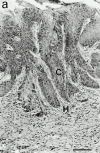
Figure 1
Figure 1
Invasive behavior of malignant mouse keratinocytes (PDVA cells), 2 wk after implantation. In WT (a), uPA−/− (b), tPA−/− (c), tPA−/−/uPA−/− (d), and uPAR−/− (e) mice, the collagen gel has been remodelled and malignant cells formed tumor sprouts intermingled with host tissue. The dotted line delineates the bottom of the collagen gel in Plg−/− (f) and PAI-1−/− mice (g). Histological sections were stained with hematoxylin and eosin. C, carcinoma cells; G, collagen gel; H, host connective tissue. Bar, 100 μm.
Figure 1
Figure 1
Invasive behavior of malignant mouse keratinocytes (PDVA cells), 2 wk after implantation. In WT (a), uPA−/− (b), tPA−/− (c), tPA−/−/uPA−/− (d), and uPAR−/− (e) mice, the collagen gel has been remodelled and malignant cells formed tumor sprouts intermingled with host tissue. The dotted line delineates the bottom of the collagen gel in Plg−/− (f) and PAI-1−/− mice (g). Histological sections were stained with hematoxylin and eosin. C, carcinoma cells; G, collagen gel; H, host connective tissue. Bar, 100 μm.
Figure 2
Figure 2
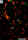
Immunofluorescence labeling of malignant keratinocytes and vessels 2 wk after implantation. PDVA cells were transplanted into WT (a), uPA−/− (b), tPA−/− (c), tPA/uPA−/− (d), uPAR−/− (e), Plg−/− (f), and PAI-1−/− (g) mice. Malignant cells were detected on cryostat sections by using antikeratin Ab (green) and vessels were detected using an anticollagen type IV Ab (red). At all times after grafting, collagen type IV labelings were codistributed with endothelial cells recognized by the anti–mouse platelet endothelial cell adhesion molecule immunostaining (data not shown). The dotted line delineates the bottom of the collagen gel, revealing not only a reduced upward migration of vessels in Plg−/− mice (e), but also that vessels remained confined beneath the collagen gel in PAI-1−/− mice (f). C, carcinoma cells; G, collagen gel; H, host connective tissue. Bar, 100 μm.
Figure 2
Figure 2
Immunofluorescence labeling of malignant keratinocytes and vessels 2 wk after implantation. PDVA cells were transplanted into WT (a), uPA−/− (b), tPA−/− (c), tPA/uPA−/− (d), uPAR−/− (e), Plg−/− (f), and PAI-1−/− (g) mice. Malignant cells were detected on cryostat sections by using antikeratin Ab (green) and vessels were detected using an anticollagen type IV Ab (red). At all times after grafting, collagen type IV labelings were codistributed with endothelial cells recognized by the anti–mouse platelet endothelial cell adhesion molecule immunostaining (data not shown). The dotted line delineates the bottom of the collagen gel, revealing not only a reduced upward migration of vessels in Plg−/− mice (e), but also that vessels remained confined beneath the collagen gel in PAI-1−/− mice (f). C, carcinoma cells; G, collagen gel; H, host connective tissue. Bar, 100 μm.
Figure 3
Figure 3
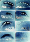
In situ zymography of PDVA cells 2 wk after transplantation into WT (a and b), uPA−/− (c and d), tPA−/− (e and f), and uPAR−/− (g and h) mice. Total PA activity was visualized as a dark zone of lysis (dark field images) after incubation for 6 h (a, c, e, and g). In the presence of 2 mM amiloride for 36 h, only tPA activity was detected (b, d, f, and h). C, carcinoma cells; H, host connective tissue. Bar, 1 mm.
Figure 4
Figure 4
Invasive behavior of malignant mouse keratinocytes (PDVA cells) 2 wk after implantation into WT mice (a) or Vn−/− mice (b). Histological sections stained with hematoxylin and eosin revealed tumor cells (C) intermingled with host cells (H) in both WT mice (a) and Vn−/− mice (b). Bar, 100 μm.

References

    1. Andreasen P.A., Kjoller L., Christensen L., Duffy M.J. The urokinase-type plasminogen activator system in cancer metastasisa review. Int. J. Cancer. 1997;72:1–22. - PubMed
    1. Bajou K., Noël A., Gerard R.D., Masson V., Brunner N., Holst-Hansen C., Skobe M., Fusenig N.E., Carmeliet P., Collen D., Foidart J.M. Absence of host plasminogen activator inhibitor 1 prevents cancer invasion and vascularization. Nat. Med. 1998;4:923–928. - PubMed
    1. Blasi F. uPA, uPAR, PAI-1key intersection of proteolytic, adhesive and chemotactic highways? Immunol. Today. 1997;18:415–417. - PubMed
    1. Brunner N., Stephens R.W., Dano K. Control of invasion and metastasis. In: Harris J.R., editor. Disease of the Breast. Lippincott; Williams & Wilkins, Philadelphia, PA: 2000. pp. 367–375.
    1. Bugge T.H., Flick M.J., Daugherty C.C., Degen J.L. Plasminogen deficiency causes severe thrombosis but is compatible with development and reproduction. Genes Dev. 1995;9:794–807. - PubMed

Publication types

MeSH terms