. 2001 Feb 19;152(4):795-808.
doi: 10.1083/jcb.152.4.795.

Rab27a regulates the peripheral distribution of melanosomes in melanocytes

Affiliations

Rab27a regulates the peripheral distribution of melanosomes in melanocytes

A N Hume et al. J Cell Biol. .

Abstract

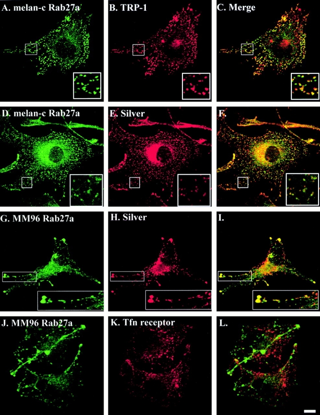
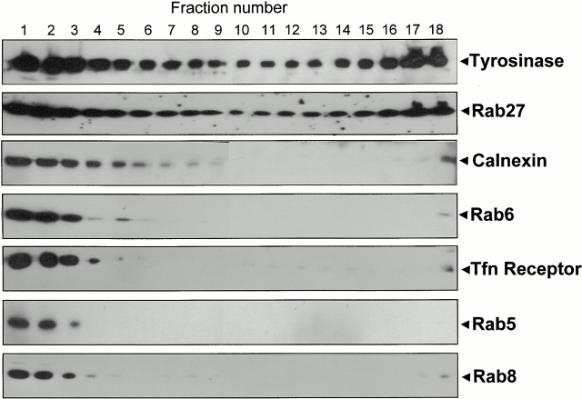
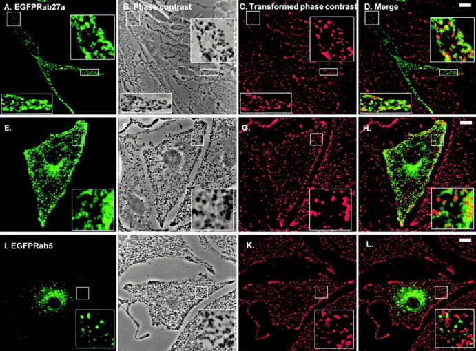
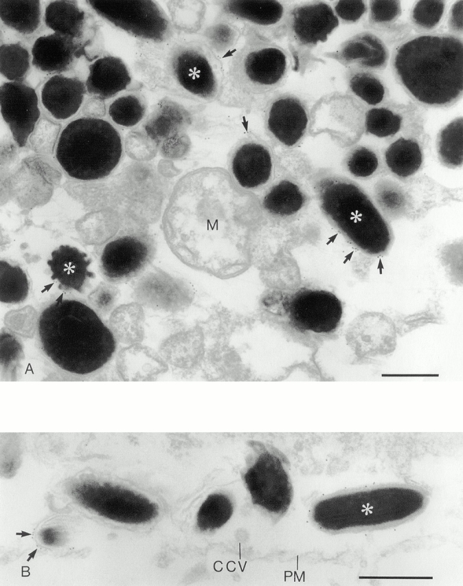
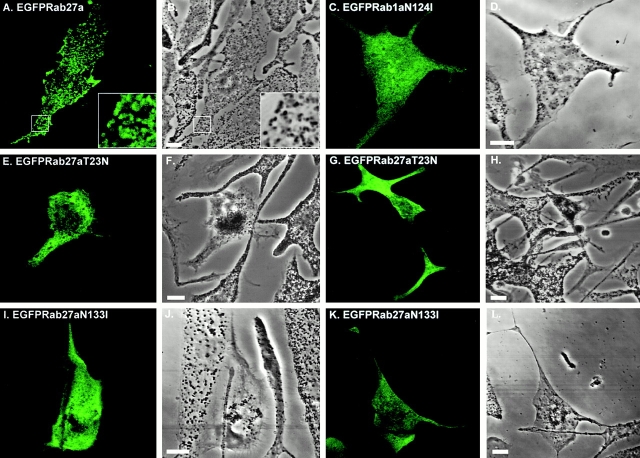
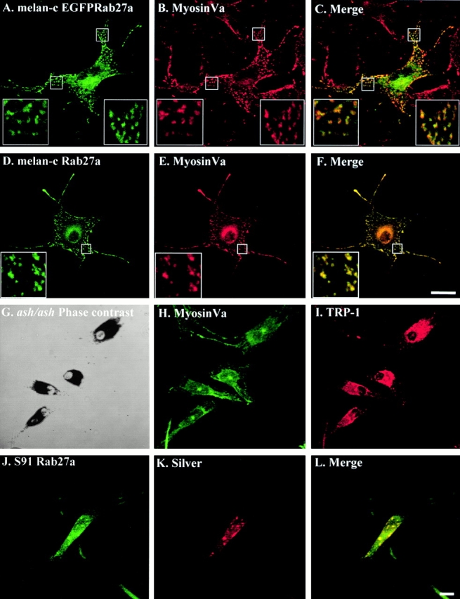
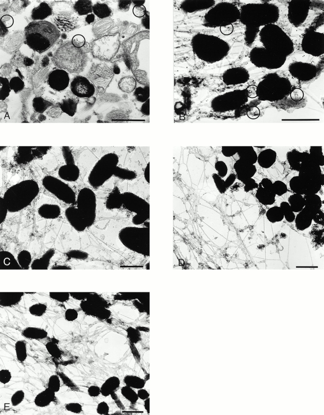
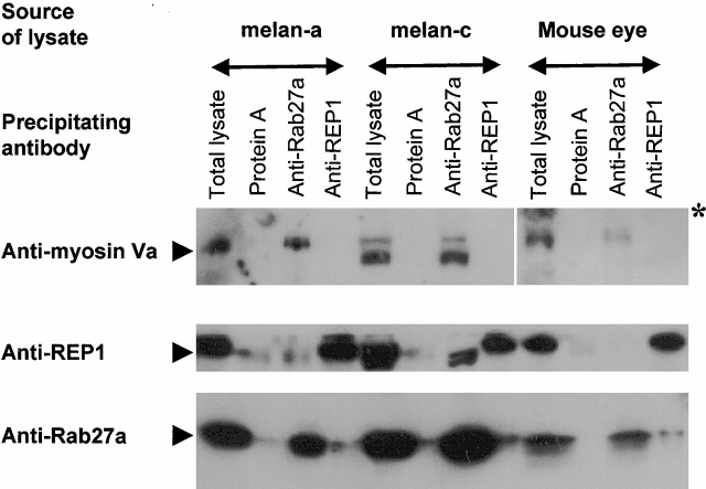
Rab GTPases are regulators of intracellular membrane traffic. We report a possible function of Rab27a, a protein implicated in several diseases, including Griscelli syndrome, choroideremia, and the Hermansky-Pudlak syndrome mouse model, gunmetal. We studied endogenous Rab27a and overexpressed enhanced GFP-Rab27a fusion protein in several cultured melanocyte and melanoma-derived cell lines. In pigmented cells, we observed that Rab27a decorates melanosomes, whereas in nonpigmented cells Rab27a colocalizes with melanosome-resident proteins. When dominant interfering Rab27a mutants were expressed in pigmented cells, we observed a redistribution of pigment granules with perinuclear clustering. This phenotype is similar to that observed by others in melanocytes derived from the ashen and dilute mutant mice, which bear mutations in the Rab27a and MyoVa loci, respectively. We also found that myosinVa coimmunoprecipitates with Rab27a in extracts from melanocytes and that both Rab27a and myosinVa colocalize on the cytoplasmic face of peripheral melanosomes in wild-type melanocytes. However, the amount of myosinVa in melanosomes from Rab27a-deficient ashen melanocytes is greatly reduced. These results, together with recent data implicating myosinVa in the peripheral capture of melanosomes, suggest that Rab27a is necessary for the recruitment of myosinVa, so allowing the peripheral retention of melanosomes in melanocytes.


Figures

Figure 1
Figure 1
Rab27a colocalizes with melanosome-resident proteins in MM96 and melan-c cells. Coverslip-grown melan-c or MM96 cells were fixed and stained using the polyclonal anti-Rab27a antibody Q142 (A, D, G, and J); monoclonal antibodies reactive to the melanosomal marker proteins TRP-1 (B) or silver (E and H); and the transferrin receptor (Tfn; panel K), an endosome marker, as described in Materials and Methods. Polyclonal antibodies were detected using Alexa 488–conjugated goat anti–rabbit secondary antibodies (A, D, G, and J); monoclonal antibodies were detected using Alexa 568–conjugated goat anti–mouse second antibodies (B, E, H, and K). Merged fluorescent signals for Rab27a and the indicated marker proteins are shown in C, F, I, and L. Bar, 10 μm. See Figure S1, available at http://www.jcb.org/cgi/content/full/152/4/795/DC1.
Figure 4
Figure 4
Subcellular fractionation of melanocyte-derived membranes. Postnuclear supernatants of melan-a cells were subjected to a 28% Percoll gradient centrifugation; fractions were then collected from the top of the gradient and analyzed by immunoblot. The lightest fraction is 1 (top of the gradient) and the densest fraction is 18 (bottom of the gradient). Antibodies used are indicated on the right. Rab5, Rab6, and Rab8 localize to early endosomes, Golgi, and TGN/plasma membrane, respectively. Calnexin, the transferrin (Tfn) receptor, and tyrosinase are membrane proteins that localize to the ER, early endosomes, and melanosomes, respectively.
Figure 2
Figure 2
Transiently overexpressed EGFP-Rab27a is associated with pigment granules in melan-a cells. Melan-a cells were transfected with pEGFP-Rab27a (A–H) or pEGFP-Rab5 (I–L) and fixed 48 h later, as described in Materials and Methods with the following modification. After transfection, cells were permeabilized for 5 min before fixation in solution 4 (80 mM K-Pipes, pH 6.8, 5 mM EGTA, 1 mM MgCl2, 0.05% saponin) in order to remove nonmembrane-associated fusion protein. A, E, and I show the distribution of the indicated overexpressed EGFP-Rab fusion protein in melan-a cells. B, F, and J are phase–contrast images of melan-a cells showing the distribution of pigment granules in transfected cells. Phase–contrast images (B, F, and J) of melan-a cells were transformed into pseudocolor (red) images, shown in C, G, and K, using Adobe Photoshop® 4.0 and then merged with the green fluorescence signal emitted by overexpressed EGFP-Rab27a or EGFP-Rab5 proteins, respectively (D, H, and L). Bars, 10 μm.
Figure 3
Figure 3
Immunoelectron microscopy of Rab27a in melan-a cells. (A) Melan-a melanocytes contain predominantly stage IV melanosomes, many of which are labeled for Rab27a. Melan-a melanocytes were permeabilized with digitonin and the anti-Rab27a antibody detected with a 10-nm gold-conjugated secondary antibody as described in Materials and Methods. The presence of representative gold particles is indicated by arrows. Mitochondria (M) are demonstrative of other organelles in the melanocyte that show a very low level of background labeling. (B) Rab27a-positive melanosomes lie just below the plasma membrane (PM) adjacent to a clathrin-coated vesicle (CCV). Bars, 0.5 μm.
Figure 5
Figure 5
Transient overexpression of dominant interfering Rab27a mutants, Rab27aT23N and Rab27aN133I, results in perinuclear clustering of pigment granules in melanocytes. Melan-a cells were transfected with pEGFP-Rab27a (A and B), pEGFP-Rab1aN124I (C and D), pEGFP-Rab27aT23N (E–H), or pEGFP-Rab27aN133I (I–L) and fixed 48 h later, as described in Materials and Methods. A, C, E, G, I, and K show the distribution of the indicated overexpressed EGFP-Rab fusion protein in melan-a cells. B, D, F, H, J, and L are phase–contrast images showing the distribution of pigment granules in these cells. Bars, 10 μm.
Figure 6
Figure 6
MyosinVa colocalizes with Rab27a on melanosomes in wild-type melanocytes, but is absent from melanosomes in Rab27a mutant ashen melanocytes. (A–F) Melan-c cells were transfected with pEGFP-Rab27a (A) and fixed 48 h later. Endogenous Rab27a in fixed melan-c cells was stained using the polyclonal anti-Rab27a antibody Q142 detected using Alexa 488–conjugated goat anti–rabbit antibodies (D). Residual bound anti-Rab27a antibody, not detected by the Alexa 488–conjugated goat anti–rabbit antibodies, was then destroyed by a second round of fixation (see Materials and Methods). Transfected or anti-Rab27a–stained cells were then stained for myosinVa using polyclonal antimyosinVa antibody detected using Alexa 568–conjugated goat anti–rabbit antibodies (B and E). (G–I) Coverslip-grown primary cultures of melanocytes derived from the murine Rab27a mutant ashen were fixed and stained with antimyosinVa polyclonal antibodies and anti–TRP-1 monoclonal antibodies which were detected using Alexa 488–conjugated goat anti–rabbit antibodies (H) and Alexa 568–conjugated goat anti–mouse antibodies (I). G is a phase–contrast image showing the perinuclear distribution of melanosomes in ashen melanocytes. (J–L) Coverslip-grown S91/Cloudman melanoma cells derived from the murine myosinVa mutant dilute were fixed and stained with polyclonal anti-Rab27a antibody and monoclonal HMB-45 antibody detected using Alexa 488–conjugated goat anti–rabbit antibodies (J) and Alexa 568–conjugated goat anti–mouse antibodies (K). C, F, and L show merged fluorescence images. Bars, 20 μm. See supplemetal figure S2, available at http://www.jcb.org/cgi/content/full/152/4/795/DC1.
Figure 7
Figure 7
Localization of myosinVa in melan-a and ashen melanocytes. MyosinVa (15-nm gold) and Rab27a (10-nm gold) were colocalized in Epon-embedded ultrathin sections (A) and whole mounts (B) of melan-a melanocytes as described in Materials and Methods. The distribution of myosinVa in melan-a melanocytes (C) was compared with the distribution in Rab27a mutant ashen melanocytes (D). Melan-a melanocytes were incubated with the secondary 15-nm gold anti–rabbit antibody as a control for specificity of the label. Negligible levels of background labeling were observed (E). Bar, 0.5 μm.
Figure 8
Figure 8
Coimmunoprecipitation of Rab27a and myosinVa. Melanocyte or mouse eye lysates were incubated with the indicated antibodies. The adsorbed material was precipitated and analyzed by immunoblot as described in Materials and Methods.

Comment in

References

    1. Allan B.B., Moyer B.D., Balch W.E. Rab1 recruitment of p115 into a cis-SNARE complexprogramming budding COPII vesicles for fusion. Science. 2000;289:444–448. - PubMed
    1. Bahadoran P., Aberdam E., Mantoux F., Busca R., Bille K., Yalman N., de Saint-Basile G., Casaroli-Marano R., Ortonne J.-P., Ballotti R. Rab27aa key to melanosome transport in human melanocytes. J. Cell Biol. 2001;152:843–849. - PMC - PubMed
    1. Bennett D.C., Cooper P.J., Hart I.R. A line of non-tumorigenic mouse melanocytes, syngeneic with the B16 melanoma and requiring a tumour promoter for growth. Int. J. Cancer. 1987;39:414–418. - PubMed
    1. Bennett D.C., Cooper P.J., Dexter T.J., Devlin L.M., Heasman J., Nester B. Cloned mouse melanocyte lines carrying the germline mutations albino and browncomplementation in culture. Development. 1989;105:379–385. - PubMed
    1. Calvo P.A., Frank D.W., Bieler B.M., Berson J.F., Marks M.S. A cytoplasmic sequence in human tyrosinase defines a second class of di-leucine-based sorting signals for late endosomal and lysosomal delivery. J. Biol. Chem. 1999;274:12780–12789. - PubMed

Publication types

MeSH terms