. 2001 Feb 19;152(4):809-24.
doi: 10.1083/jcb.152.4.809.

Distinct protein sorting and localization to premelanosomes, melanosomes, and lysosomes in pigmented melanocytic cells

Affiliations

Distinct protein sorting and localization to premelanosomes, melanosomes, and lysosomes in pigmented melanocytic cells

G Raposo et al. J Cell Biol. .

Abstract

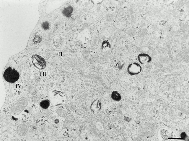
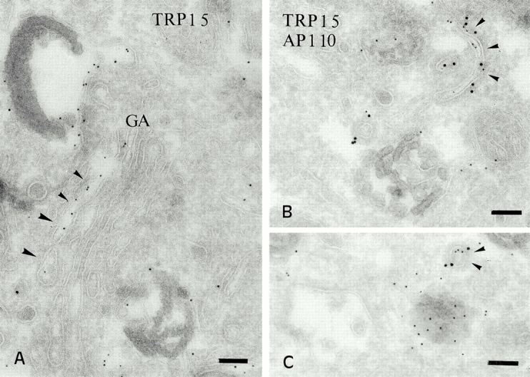
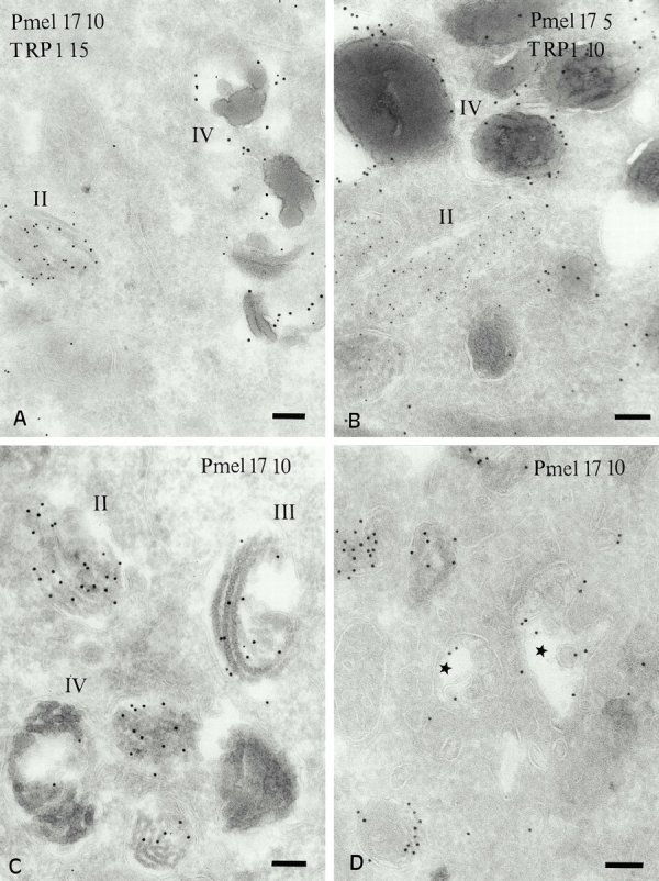
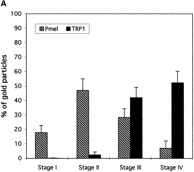
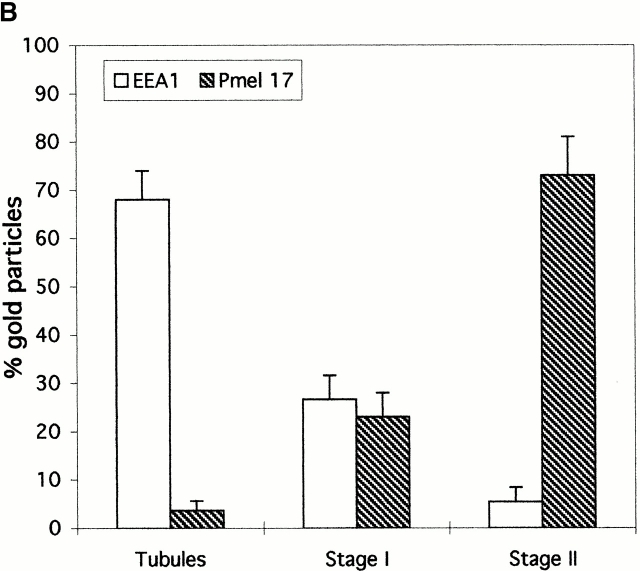
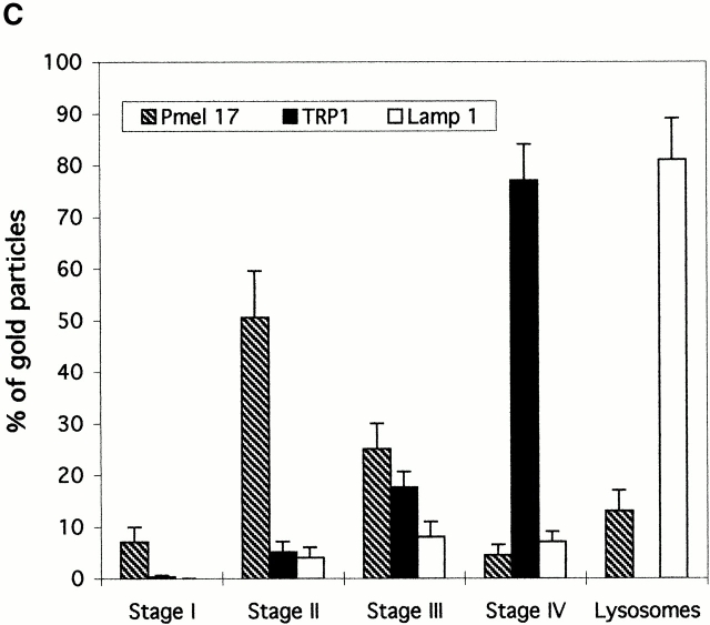
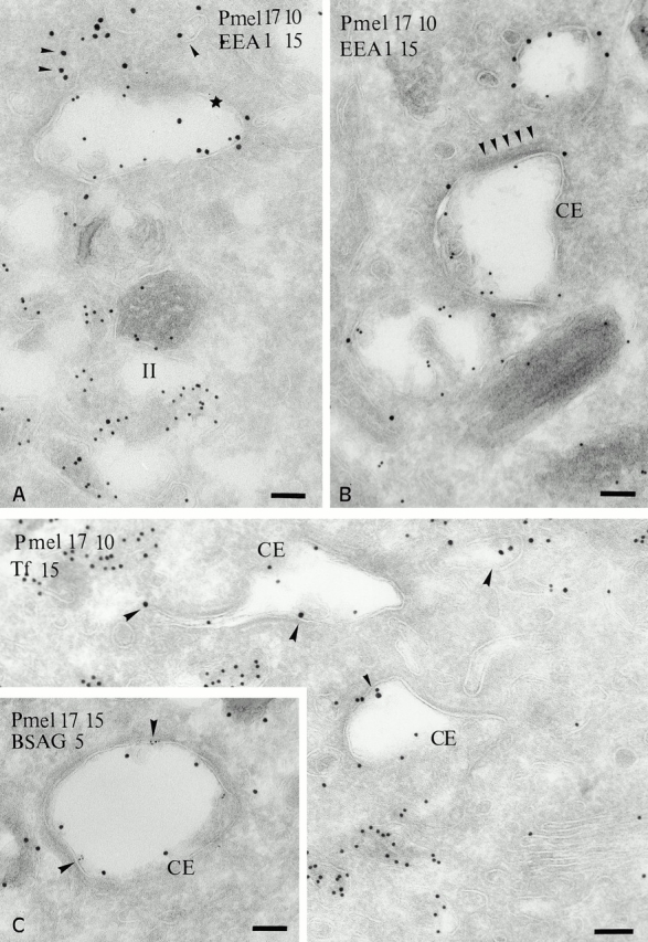
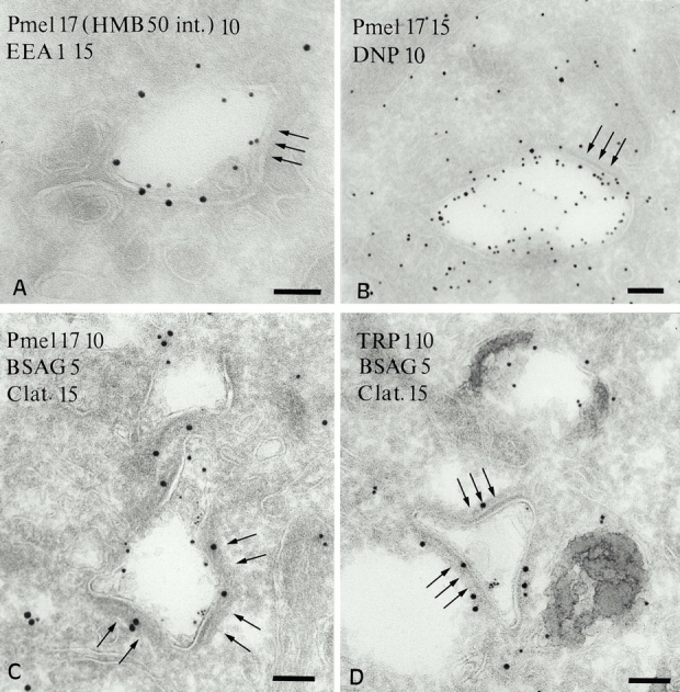
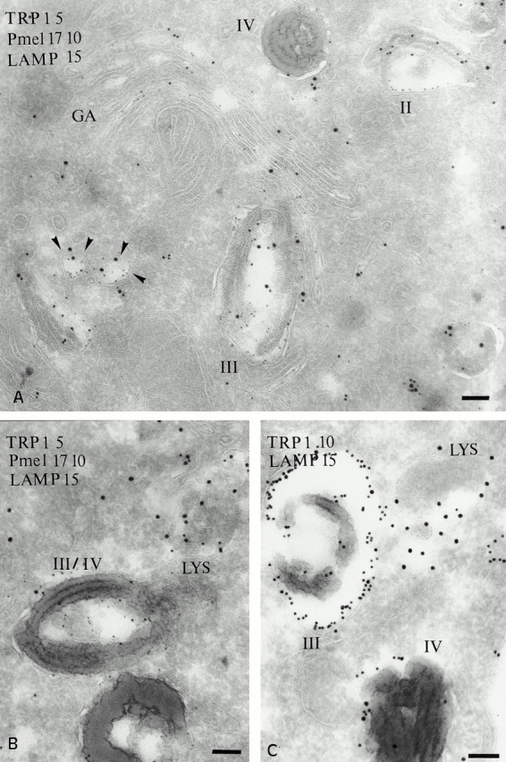
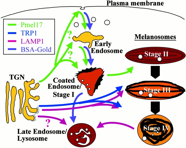
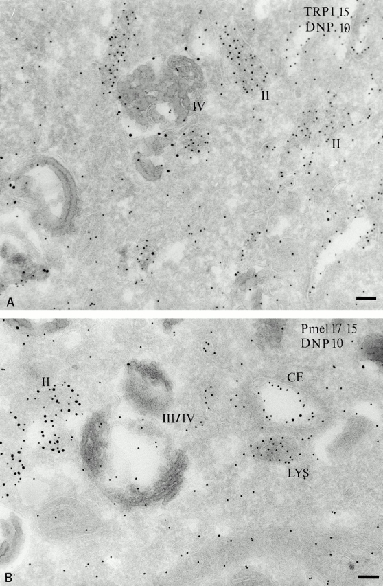
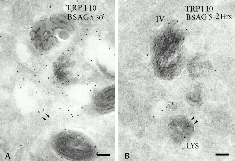
Melanosomes and premelanosomes are lysosome-related organelles with a unique structure and cohort of resident proteins. We have positioned these organelles relative to endosomes and lysosomes in pigmented melanoma cells and melanocytes. Melanosome resident proteins Pmel17 and TRP1 localized to separate vesicular structures that were distinct from those enriched in lysosomal proteins. In immunogold-labeled ultrathin cryosections, Pmel17 was most enriched along the intralumenal striations of premelanosomes. Increased pigmentation was accompanied by a decrease in Pmel17 and by an increase in TRP1 in the limiting membrane. Both proteins were largely excluded from lysosomal compartments enriched in LAMP1 and cathepsin D. By kinetic analysis of fluid phase uptake and immunogold labeling, premelanosomal proteins segregated from endocytic markers within an unusual endosomal compartment. This compartment contained Pmel17, was accessed by BSA-gold after 15 min, was acidic, and displayed a cytoplasmic planar coat that contained clathrin. Our results indicate that premelanosomes and melanosomes represent a distinct lineage of organelles, separable from conventional endosomes and lysosomes within pigmented cells. Furthermore, they implicate an unusual clathrin-coated endosomal compartment as a site from which proteins destined for premelanosomes and lysosomes are sorted.


Figures

Figure 1
Figure 1
Ultrastructure of MNT-1 cells. IEM analysis of ultrathin sections of epon-embedded MNT-1 cells reveal numerous melanosomes at different stages of maturation throughout the cytoplasm. Structures corresponding to stages I–IV melanosomes are indicated. Bar, 400 nm.
Figure 3
Figure 3
Distribution of TRP1 in the Golgi and associated vesicles. (A) Ultrathin cryosections of MNT-1 cells were labeled with anti-TRP1 and PAG 5. TRP1 is often enriched in one side of the Golgi apparatus. (B and C) Double labeling of MNT-1 cells for TRP1 (PAG 5) and γ-adaptin (PAG 10). TRP1 positive tubules and vesicles are labeled for γ-adaptin (arrowheads). Bars, 100 nm.
Figure 2
Figure 2
Distribution of Pmel17 and TRP1 in MNT-1 cells and primary melanocytes. Ultrathin cryosections of MNT-1 cells (A) or cultured primary melanocytes derived from a highly pigmented nevus (B) were double immunogold labeled with HMB50 (anti-Pmel17) and TA99 (anti-TRP1). (C and D) MNT-1 cells were immunogold labeled with the anti-Pmel17 mAbs HMB50 and HMB45, respectively. (A) Labeling for Pmel17 (PAG 10) in internal striations of stage II melanosomes, and labeling for TRP1 (PAG 15) in the limiting membrane of a stage IV melanosome. (B) In primary melanocytes, Pmel17 (PAG 5) is enriched in the lumen of a premelanosome, whereas TRP1 (PAG 10) localizes mainly to mature melanosomes (IV). (C) Labeling for Pmel17 (PAG 10) decreases during progression from stage II to IV. Note the deposits of melanin over the internal striations in the stage III melanosome. (D) Pmel17 (PAG 10) is labeled in compartments morphologically similar to multivesicular endosomes (stars). Bars, 100 nm.
Figure 7
Figure 7
Quantitative analysis of the distribution of Pmel17 relative to TRP1, EEA1, and LAMP1 in MNT-1 cells. Gold particles labeling either Pmel17 or (A) TRP1, (B) EEA1, or (C) TRP1 or LAMP1 were counted in the different endosomal, melanosomal, or lysosomal compartments in double- (A and B) or triple- (C) labeled ultrathin cryosections of MNT-1 cells. For each set, 30 cell profiles were counted in each of two experiments. Results represent a percentage of the total number of counted gold particles for each marker in the distinct morphologically defined compartments. Only the compartments shown in each experiment were taken into account.
Figure 7
Figure 7
Quantitative analysis of the distribution of Pmel17 relative to TRP1, EEA1, and LAMP1 in MNT-1 cells. Gold particles labeling either Pmel17 or (A) TRP1, (B) EEA1, or (C) TRP1 or LAMP1 were counted in the different endosomal, melanosomal, or lysosomal compartments in double- (A and B) or triple- (C) labeled ultrathin cryosections of MNT-1 cells. For each set, 30 cell profiles were counted in each of two experiments. Results represent a percentage of the total number of counted gold particles for each marker in the distinct morphologically defined compartments. Only the compartments shown in each experiment were taken into account.
Figure 4
Figure 4
Localization of Pmel17 relative to EEA1 and endocytic tracers. (A and B) Ultrathin cryosections of MNT-1 cells were double labeled with HMB50 (anti-Pmel17; PAG 10) and anti-EEA1 (PAG 15). (A) EEA1 is detected in small vesicles (arrowheads) and on the limiting membrane of a compartment that also contains Pmel17 (star). Note that labeling for Pmel17 and EEA1 is segregated (B) EEA1 (PAG 15) is enriched in electron lucent vesicular compartments that are poorly labeled for Pmel17 (PAG 10). Note the unusual morphology of the endosomal compartments containing the bulk of Pmel17 (CE), which are poorly labeled for EEA1 and display on their cytosolic side an electron dense coat (arrowheads). (C) Cryosection of MNT-1 cells exposed to Tf-FITC for 20 min at 37°C, labeled with anti-FITC antibodies and PAG 15. Tf-FITC is present in small tubulovesicular structures (arrowheads) and in Pmel17-positive (PAG 10) coated endosomes (CE). (Inset in C) MNT-1 cells pulsed with BSAG for 5 min and chased for 10 min. BSAG (5 nm) at this time point is mainly detected in the Pmel17-positive coated endosome (CE). Pmel17 was labeled with HMB50 and PAG 15. Bars, 100 nm.
Figure 5
Figure 5
Characterization of the coated endosome in MNT-1 cells. (A) HMB50 (anti-Pmel17) was bound to the cell surface at 4°C and internalized for 30 min at 37°C. Cryosections were labeled with rabbit anti–mouse IgG (PAG 10) to visualize internalized mAb and anti-EEA1 (PAG 15). Internalized Pmel17 accesses the EEA1-positive–coated endosome (arrows) during this time period. (B) Cells were incubated with DAMP for 30 min. Accumulated DAMP was detected with anti-DNP antibodies (PAG 10); Pmel17 was detected with HMB50 (PAG 15). Note the accumulation of DAMP in the coated endosome, indicating that it is acidic. (C) The coated endosome containing Pmel17 (PAG 10) and BSAG (5 nm) internalized for 15 min is labeled on its cytosolic side with an anticlathrin antibody (PAG 15; arrows). (D) TRP1 (PAG 10) is present in mature melanosomes but is not consistently detected in the clathrin-positive coated endosome accessed by BSAG after 15 min (arrows). Bars, 100 nm.
Figure 6
Figure 6
Localization of Pmel17 and TRP1 relative to the lysosomal protein LAMP1. (A and B) Ultrathin cryosections of MNT-1 cells were triple immunogold labeled for LAMP1 (PAG 15), Pmel17 (PAG 10) and TRP 1 (PAG 5). (A) Overview of the Golgi area. Stage II premelanosomes (II) displaying characteristic Pmel17-positive striations do not contain LAMP1. TRP1 is detected in the Golgi apparatus but mainly restricted to the trans-side. Vesicles containing both TRP1 and LAMP1 (arrowheads) are present near the TGN and melanosomes and to some extent in stage III/IV melanosomes (III). (B) Stage III/IV melanosomes (III/IV) are poorly labeled for LAMP1. LAMP1 is highly enriched in electron-dense compartments distinct from melanosomes (LYS). (C) Ultrathin cryosections of MNT-1 cells were double labeled with mAbs to TRP1 (PAG 10) and LAMP1 (PAG 15). TRP1 is present on the limiting membrane of stage III and IV melanosomes. LAMP1 is also detected in the limiting membrane of the mature melanosomes, but the majority of the labeling is observed in less electron-dense lysosomal compartments (LYS). Bars, 100 nm.
Figure 10
Figure 10
Model for protein sorting steps within the endocytic and melanosomal systems in pigmented melanocytic cells. Schematic representation of the endocytic and melanosomal organelles and a working model for the sorting pathways for proteins distributed among them. We propose that Pmel17 (green) is transported to stage II premelanosomes via early endosomes and the stage I premelanosome/coated endosome; transport to the coated endosome may also be direct from the TGN. In contrast, TRP1 (blue) is proposed to be targeted directly from the TGN to a preestablished stage II or III melanosome; continual transport of TRP1 and tyrosinase results in maturation, indicated by broad arrows, of the premelanosome to the stage III and IV melanosome. The deposition of black melanin in these compartments is schematized. Internalized fluid phase markers such as BSAG (violet) are targeted to lysosomes from the coated endosome after passage through early endosomes. LAMP1 (rose) may be sorted to late endosomes/lysosomes directly from the TGN or indirectly via melanosomes; at least a fraction of LAMP1 progresses through this latter pathway. Degree of acidity is indicated by yellow/brown color, with yellow being least acidic and dark brown being most acidic.
Figure 8
Figure 8
Acidity of melanosomal compartments probed with DAMP. MNT-1 cells were incubated with DAMP for 30 min at 37°C before fixation and processing. (A) DAMP, visualized with anti-DNP antibodies (PAG 10), accumulates primarily in stage II melanosomes displaying characteristic striations. TRP1-positive stage IV melanosomes show less labeling. (B) Double immunogold localization of DAMP (PAG 10) and Pmel17 (PAG 15). DAMP is distributed over the coated endosome, stage II and IV melanosomes, and lysosomes. Bars, 100 nm.
Figure 9
Figure 9
Mature melanosomes in MNT-1 cells are not accessed by endocytic tracers. (A) BSAG (5-nm) was internalized for 30 min. BSAG is present in coated endosomes and multivesicular bodies (arrowheads) situated near mature melanosomes labeled for TRP1 (PAG 10) but does not label the melanosomes. (B) BSAG (5-nm) internalized for 2 h accumulates in electron-dense lysosomes but not in TRP1-positive (PAG 10) melanosomes (IV). Bars, 100 nm.

Comment in

References

    1. Ahle S., Mann A., Eichelsbacher U., Ungewickell E. Structural relationships between clathrin assembly proteins from the Golgi and the plasma membrane. EMBO (Eur. Mol. Biol. Organ.) J. 1988;7:919–929. - PMC - PubMed
    1. Anderson R.G., Falck J.R., Goldstein J.L., Brown M.S. Visualization of acidic organelles in intact cells by electron microscopy. Proc. Natl. Acad. Sci. USA. 1984;81:4838–4842. - PMC - PubMed
    1. Arvan P., Castle D. Sorting and storage during secretory granule biogenesislooking backward and looking forward. Biochem. J. 1998;332:593–610. - PMC - PubMed
    1. Bhatnagar V., Anjaiah S., Puri N., Darshanam N.A., Ramaiah A. pH of melanosomes of B16 murine melanoma is acidicits physiological importance in the regulation of melanin biosynthesis. Arch. Biochem. Biophys. 1993;307:183–192. - PubMed
    1. Blagoveshchenskaya A.D., Hewitt E.W., Cutler D.F. Di-leucine signals mediate targeting of tyrosinase and synaptotagmin to synaptic-like microvesicles within PC12 cells. Mol. Biol. Cell. 1999;10:3979–3990. - PMC - PubMed

Publication types