. 2001 Feb 19;152(4):843-50.
doi: 10.1083/jcb.152.4.843.

Rab27a: A key to melanosome transport in human melanocytes

Affiliations

Rab27a: A key to melanosome transport in human melanocytes

P Bahadoran et al. J Cell Biol. .

Abstract

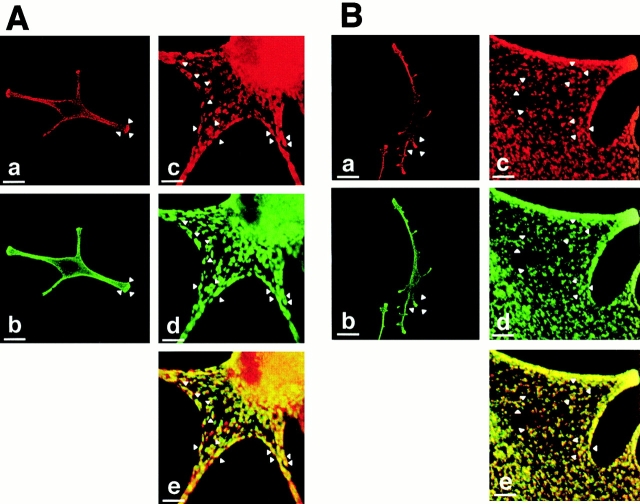
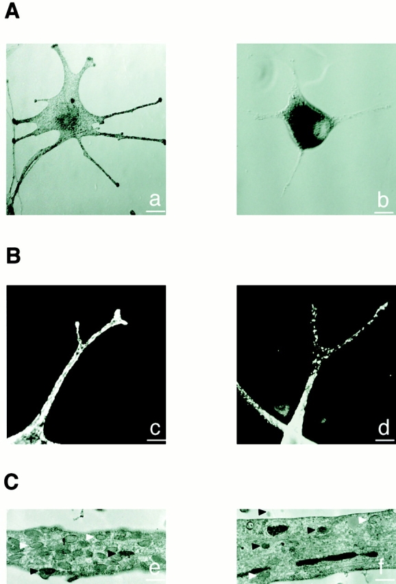
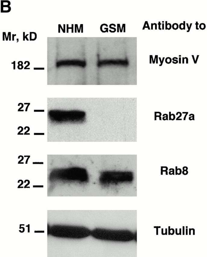
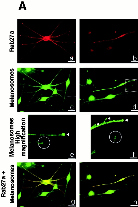
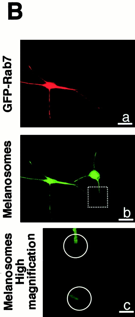
Normal pigmentation depends on the uniform distribution of melanin-containing vesicles, the melanosomes, in the epidermis. Griscelli syndrome (GS) is a rare autosomal recessive disease, characterized by an immune deficiency and a partial albinism that has been ascribed to an abnormal melanosome distribution. GS maps to 15q21 and was first associated with mutations in the myosin-V gene. However, it was demonstrated recently that GS can also be caused by a mutation in the Rab27a gene. These observations prompted us to investigate the role of Rab27a in melanosome transport. Using immunofluorescence and immunoelectron microscopy studies, we show that in normal melanocytes Rab27a colocalizes with melanosomes. In melanocytes isolated from a patient with GS, we show an abnormal melanosome distribution and a lack of Rab27a expression. Finally, reexpression of Rab27a in GS melanocytes restored melanosome transport to dendrite tips, leading to a phenotypic reversion of the diseased cells. These results identify Rab27a as a key component of vesicle transport machinery in melanocytes.


Figures

Figure 1
Figure 1
Rab27a colocalizes with melanosomes and myosin-V in normal human melanocytes. (A) Immunofluorescence microscopy of normal human melanocytes labeled with a monoclonal antibody to Rab27a (a) and with a polyclonal antibody to TRP-1 (b). Confocal immunofluorescence microscopy of normal human melanocytes labeled with a monoclonal antibody to Rab27a (c) and with a polyclonal antibody to TRP-1 (d). Image overlay (e). Arrowheads indicate the colocalization of Rab27a and melanosomes. (B) Immunofluorescence microscopy of normal human melanocytes labeled with a monoclonal antibody to Rab27a (a) and with a polyclonal antibody to myosin-V (b). Confocal microscopy images of normal human melanocytes labeled with a monoclonal antibody to Rab27a (c) and a polyclonal antibody to myosin-V (d). Image overlay (e). Arrowheads indicate the colocalization of Rab27a and myosin-V. (C) Immunogold labeling of normal human melanocytes with the anti-Rab27a antibody (15-nm gold particles) (a). Immunogold labeling of normal human melanocytes with the anti-Rab27a antibody (15-nm gold particles) and the anti–TRP-1 antibody (5-nm gold particles) (b, c, and d). Black arrows indicate Rab27a and white arrows indicate TRP-1. Bars: (A, a and b) 30 μm; (A, c–e) 3 μm; (B, a and b) 30 μm; (B, c–e) 3 μm; (C, a) 0.6 μm; (C, b–d) 0.2 μm.
Figure 1
Figure 1
Rab27a colocalizes with melanosomes and myosin-V in normal human melanocytes. (A) Immunofluorescence microscopy of normal human melanocytes labeled with a monoclonal antibody to Rab27a (a) and with a polyclonal antibody to TRP-1 (b). Confocal immunofluorescence microscopy of normal human melanocytes labeled with a monoclonal antibody to Rab27a (c) and with a polyclonal antibody to TRP-1 (d). Image overlay (e). Arrowheads indicate the colocalization of Rab27a and melanosomes. (B) Immunofluorescence microscopy of normal human melanocytes labeled with a monoclonal antibody to Rab27a (a) and with a polyclonal antibody to myosin-V (b). Confocal microscopy images of normal human melanocytes labeled with a monoclonal antibody to Rab27a (c) and a polyclonal antibody to myosin-V (d). Image overlay (e). Arrowheads indicate the colocalization of Rab27a and myosin-V. (C) Immunogold labeling of normal human melanocytes with the anti-Rab27a antibody (15-nm gold particles) (a). Immunogold labeling of normal human melanocytes with the anti-Rab27a antibody (15-nm gold particles) and the anti–TRP-1 antibody (5-nm gold particles) (b, c, and d). Black arrows indicate Rab27a and white arrows indicate TRP-1. Bars: (A, a and b) 30 μm; (A, c–e) 3 μm; (B, a and b) 30 μm; (B, c–e) 3 μm; (C, a) 0.6 μm; (C, b–d) 0.2 μm.
Figure 2
Figure 2
Melanosome distribution is markedly impaired in GS melanocytes. (A) Phase–contrast microscopy images of a normal (a) and GS (b) melanocyte. (B) Confocal immunofluorescence microscopy images of a normal (c) and GS (d) melanocyte labeled for melanosomes with an anti–TRP-1 antibody. (C) Electron microscopy images of a dendritic extension of a normal (e) and GS (f) melanocyte. Note that despite the reduction of melanosome number, both partially (white arrowheads) and fully pigmented (black arrowheads) melanosomes are found in the dendrite of the GS melanocyte. Bars: (A) 25 μm; (B) 5 μm; (C) 1 μm.
Figure 3
Figure 3
Absence of Rab27a expression in GS melanocytes. (A) Immunofluorescence labeling of myosin-V in normal (a) and GS (b) melanocytes, and of Rab27a in normal (c) and GS (d) melanocytes. For Rab27a labeling, Hoechst staining was used to visualize the nuclei. (B) Western blot analysis. Membranes were blotted with a polyclonal antimyosin-V antibody (top). Membranes were blotted first with a monoclonal anti-Rab27a antibody (middle). After stripping, membranes were reblotted with a monoclonal anti-Rab8 antibody. Membranes were blotted with a monoclonal antitubulin antibody to control the equal amount of protein in each lane (bottom). Bars, 30 μm.
Figure 3
Figure 3
Absence of Rab27a expression in GS melanocytes. (A) Immunofluorescence labeling of myosin-V in normal (a) and GS (b) melanocytes, and of Rab27a in normal (c) and GS (d) melanocytes. For Rab27a labeling, Hoechst staining was used to visualize the nuclei. (B) Western blot analysis. Membranes were blotted with a polyclonal antimyosin-V antibody (top). Membranes were blotted first with a monoclonal anti-Rab27a antibody (middle). After stripping, membranes were reblotted with a monoclonal anti-Rab8 antibody. Membranes were blotted with a monoclonal antitubulin antibody to control the equal amount of protein in each lane (bottom). Bars, 30 μm.
Figure 4
Figure 4
Reexpression of Rab27a restores melanosome transport to dendrite tips in GS melanocytes. Immunofluorescence labeling of two representative fields of GS melanocytes transfected with the normal Rab27a cDNA. The cells were labeled with a monoclonal antibody to Rab27a (a and b) and the melanosomes with a polyclonal antibody to TRP1 (c–f). Higher magnification of dendrite tips (e and f) clearly shows the accumulation of melanosomes at dendrite tips of transfected cells (white arrowheads) contrasting with the vacuity of dendrite extremities in untransfected GS melanocytes (white circles). Image overlay (g and h) shows the colocalization of transfected Rab27a with melanosomes. (B) Representative field of GS melanocytes transfected with GFP-Rab7 cDNA (shown in red by artificial color) (a). The cells were labeled for melanosomes with a polyclonal antibody to TRP-1 (b). Higher magnification of dendrite tips (c) clearly shows the absence of melanosome accumulation at dendrite tips in both untransfected and transfected melanocytes (b and c). Bars: (A, a–d, g, and h) 30 μm; (A, e and f) 8 μm; (B, a and b) 30 μm; (B, c) 8 μm.
Figure 4
Figure 4
Reexpression of Rab27a restores melanosome transport to dendrite tips in GS melanocytes. Immunofluorescence labeling of two representative fields of GS melanocytes transfected with the normal Rab27a cDNA. The cells were labeled with a monoclonal antibody to Rab27a (a and b) and the melanosomes with a polyclonal antibody to TRP1 (c–f). Higher magnification of dendrite tips (e and f) clearly shows the accumulation of melanosomes at dendrite tips of transfected cells (white arrowheads) contrasting with the vacuity of dendrite extremities in untransfected GS melanocytes (white circles). Image overlay (g and h) shows the colocalization of transfected Rab27a with melanosomes. (B) Representative field of GS melanocytes transfected with GFP-Rab7 cDNA (shown in red by artificial color) (a). The cells were labeled for melanosomes with a polyclonal antibody to TRP-1 (b). Higher magnification of dendrite tips (c) clearly shows the absence of melanosome accumulation at dendrite tips in both untransfected and transfected melanocytes (b and c). Bars: (A, a–d, g, and h) 30 μm; (A, e and f) 8 μm; (B, a and b) 30 μm; (B, c) 8 μm.

Comment in

References

    1. Aberdam E., Romero C., Ortonne J.P. Repeated UVB irradiations do not have the same potential to promote stimulation of melanogenesis in cultured normal human melanocytes. J. Cell Sci. 1993;106:1015–1022. - PubMed
    1. Buscà R., Martinez M., Vilella E., Pognonec P., Deeb S., Auwerw J., Reina M., Vilaro S. The mutation Gly142-->Glu in human lipoprotein lipase produces a missorted protein that is diverted to lysosomes. J. Biol. Chem. 1996;271:2139–2146. - PubMed
    1. Cheney R.E., O'Shea M.K., Heuser J.E., Coelho M.V., Wolenski J.S., Espreafico E.M., Forscher P., Larson R.E., Mooseker M.S. Brain myosin-V is a two-headed unconventional myosin with motor activity. Cell. 1993;75:13–23. - PubMed
    1. Dufourcq-Lagelouse R., Pastural E., Barrat F.J., Feldmann J., Le Deist F., Fischer A., De Saint Basile G. Genetic basis of hemophagocytic lymphohistiocytosis syndrome. Int. J. Mol. Med. 1999;4:127–133. - PubMed
    1. Echard A., Jollivet F., Martinez O., Lacapere J.J., Rousselet A., Janoueix-Lerosey I., Goud B. Interaction of a Golgi-associated kinesin-like protein with Rab6. Science. 1998;279:580–585. - PubMed

Publication types

MeSH terms