. 2001 Apr 1;29(7):1491-506.
doi: 10.1093/nar/29.7.1491.

DNA methyltransferases of the cyanobacterium Anabaena PCC 7120

Affiliations

DNA methyltransferases of the cyanobacterium Anabaena PCC 7120

A V Matveyev et al. Nucleic Acids Res. .

Abstract

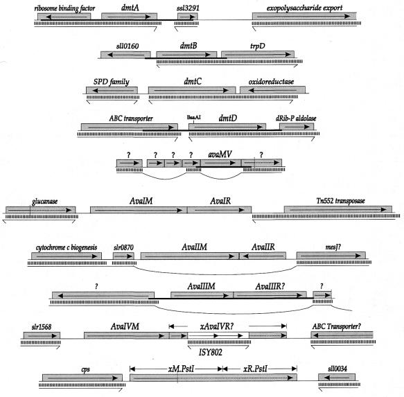
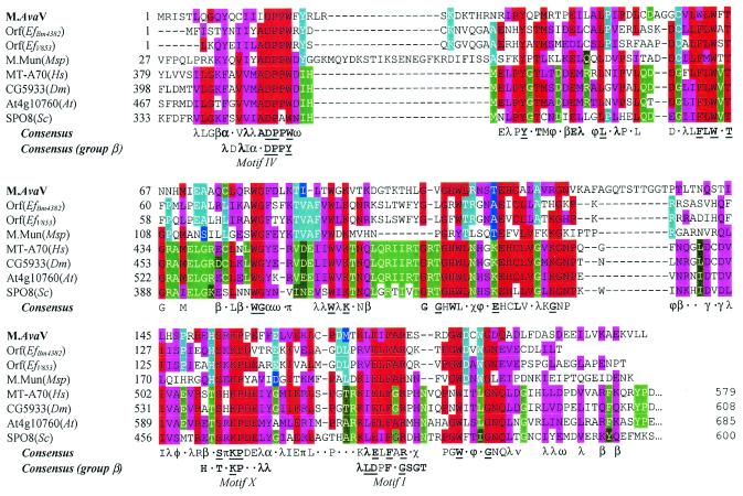
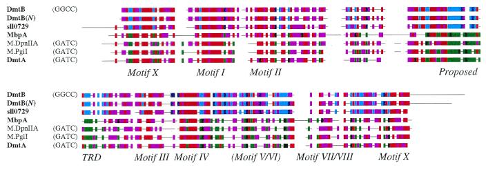
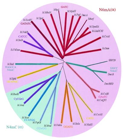
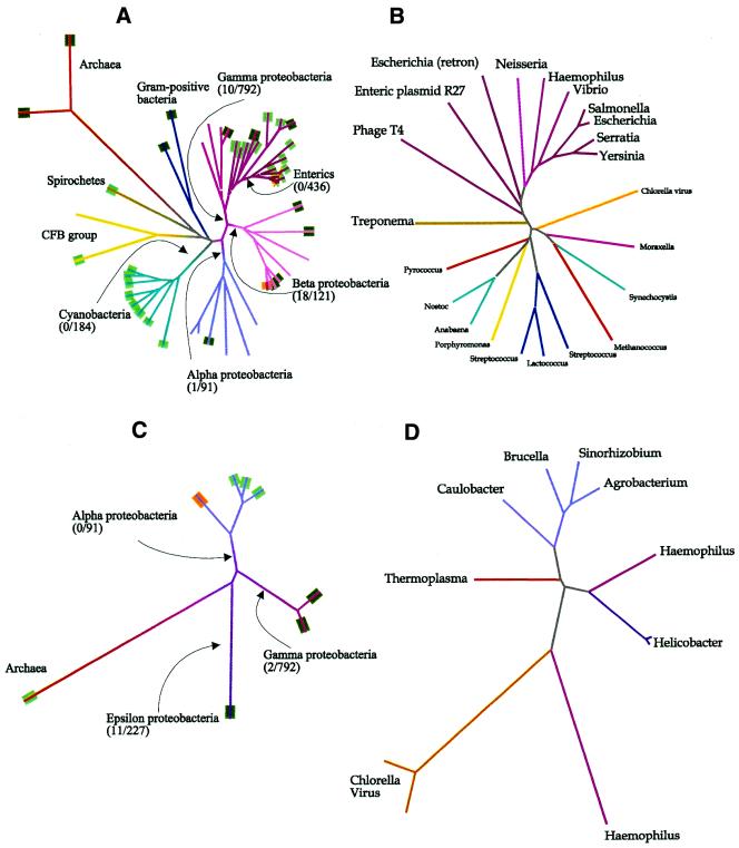
From the characterization of enzyme activities and the analysis of genomic sequences, the complement of DNA methyltransferases (MTases) possessed by the cyanobacterium ANABAENA PCC 7120 has been deduced. ANABAENA has nine DNA MTases. Four are associated with Type II restriction enzymes (AVAI, AVAII, AVAIII and the newly recognized inactive AVAIV), and five are not. Of the latter, four may be classified as solitary MTases, those whose function lies outside of a restriction/modification system. The group is defined here based on biochemical and genetic characteristics. The four solitary MTases, DmtA/M.AVAVI, DmtB/M.AVAVII, DmtC/M. AVAVIII and DmtD/M.AVAIX, methylate at GATC, GGCC, CGATCG and rCCGGy, respectively. DmtB methylates cytosines at the N4 position, but its sequence is more similar to N6-adenine MTases than to cytosine-specific enzymes, indicating that it may have evolved from the former. The solitary MTases, appear to be of ancient origin within cyanobacteria, while the restriction MTases appear to have arrived by recent horizontal transfer as did five now inactive Type I restriction systems. One Mtase, M.AVAV, cannot reliably be classified as either a solitary or restriction MTase. It is structurally unusual and along with a few proteins of prokaryotic and eukaryotic origin defines a structural class of MTases distinct from all previously described.


Figures

Figure 1
Figure 1
Genetic context surrounding genes encoding or potentially encoding DNA MTases of Anabaena. Gray boxes represent ORFs, with the direction of translation shown. The white box indicates an insertion sequence. Vertical dotted lines indicate the point at which a frameshift occurs. Thick horizontal lines indicate the region that was cloned. Thin horizontal lines separated from the Anabaena genes by vertical bars indicate that a contiguous DNA sequence from N.punctiforme exhibits sequence similarity in that region. Horizontal lines with bent ends represent DNA sequences that continue without similarity to the Anabaena sequence. Loops indicate interpolations of sequences without similarity to the Anabaena sequence. Genes with names preceded by ‘x’ are defective by reason of a frameshift, nonsense codon or insertion sequence.
Figure 2
Figure 2
Alignment of M.AvaV with known and putative DNA and RNA MTases. Sequences listed in Table 1 (except the mouse sequence, which is substantially the same as the human sequence) were aligned as described in Materials and Methods. For each position, amino acids substantially conserved over the entire group are colored red and similar amino acids colored magenta. Identical amino acids within the bacterial subgroup (first four sequences) are colored cyan and similar amino acids dark blue. Identical amino acids within the eukaryotic subgroup (last four sequences) are colored bright green and similar amino acids dark green. A consensus sequence over the entire group is shown below the alignment and for the corresponding region of Group β N6mA DNA MTases (10). A letter is shown if >50% of the sequences have the same amino acid residue or class of amino acids at the given position. The letter is in bold type if the frequency is >75% and underscored if the position is invariant. α, aromatic (FHWY); β, big hydrophilic (EKQR); λ, leucine family (ILMV); ν, negative (DE); π, positive (KR); γ, glutamate family (EQDK); χ, (NHY); φ, (ANST); ω, aspartate family (END).
Figure 3
Figure 3
Alignment of DmtB and DmtA with representative Group α Nm6A/Nm4C DNA MTases. Names of cyanobacterial sequences are shown in bold. Coloring conventions are as described in the legend to Figure 2, with the cyan/dark blue and green/dark green groups representing DNA MTases of proven and putative GGCC-specificity and GATC-specificity, respectively. The motifs (including the TRD, target recognition domain) are those described by Tran et al. (15).
Figure 4
Figure 4
Dendrogram of all available sequences of N4 and N6 MTases of the α group. The sequences compared consisted of the N- and C-termini, with the central TRD removed. The TRD was liberally defined as the block corresponding to residues 77 through 172 according to M.DpnII coordinates (15). The branches of all N6mA MTases are some hue of red and of all N4mC MTase some hue of blue. Thin lines indicate that the target sequence of the enzyme has not been established by biochemical criteria. Besides those sequences listed in Table 1, the following were found through the indicated accession nos: M.AvaI (CAA66984), M.BsoBI (CAA66933), M.CviAII (S27901), M.CviBI (Q01511), M.EcDam (P00475), M.EcoRV (P04393), M.EcoT4Dam (X17641), M.FokI (P14871), M.MboIA (P34720), M.MvaI (P14244), M.NcoI (AAC23514), M.NlaIII, M.PspGI (AAC67523), M.SphI (AAB40377) and M.TpaI (AAB82782). M.FokI and M.StsI consist of two separable MTases that are fused as a single polypeptide. The N-terminal portions (M.FokIa and M.StsIa) recognize one asymmetric target and the C-terminal portions (M.FokIb and M.StsIb) recognize the other.
Figure 5
Figure 5
Relationship amongst enzymes functionally similar to known solitary MTases, Dam and CcrM. Aligned 16S rRNA sequences were obtained from the Ribosomal Database Project (http://www.cme.msu.edu/RDP/html/index.html), and only the part of the alignment common to all was used in constructing the tree. (A) Dendrogram of 16S rRNA sequences from organisms that have been tested for GATC-specific adenine methylation (5) or that possess sequenced GATC-specific MTases of type N6mA(α). The number of restriction endonucleases known to recognize GATC is given followed by the total number of known endonucleases, for all bacteria beyond the indicated branch point. Lines are colored according to taxonomic groups. Tags at the end of branches are green if GATC-specific methylation has been demonstrated, dark green if the strain possesses a GATC-specific MTase and orange if the strain possesses a proven solitary GATC-specific MTase. (B) Dendrogram of all GATC-specific MTases of type N6mA(α) for which sequences are available. The lines are colored as in (A). The source of the sequences are given in the legend to Figure 4, except for Dam from Yersinia pseudotuberculosis (AAG23175), Dam from Serratia marcescens (P45454), Dam from S.typhimurium (P55893), gene from V.cholerae O395 (AAG23174), gene from H.influenzae Rf (P44431), Dam from Neisseria meningitidis BF13 (AAD34292), gene from E.coli retron Ec67 (P21311) and gene from enteric plasmid R27 (AAF69879). (C) Dendrogram of 16S rRNA sequences from organisms that possess sequenced GAnTC-specific MTases. The conventions are analogous to those described in (A). (D) Dendrogram of all GAnTC-specific MTases for which sequences are available. The lines are colored as in (C). The source of the sequences are (reading counterclockwise from Agrobacterium): gene from Agrobacterium tumefaciens C58 (Brad Goodner, personal communication), gene from Sinorhizobium meliloti 1021 (AAB71350), M.BabI (O30570), CcrM (Q45971), gene from Thermoplasma acidophilum DSM1728 (CAC12293), M.CviBI (Q01511), M.CviQVI (AAC03126), M.HhaII (P00473), M.HpyAIV (H64688), M.Hpy99IX (F71827) and M.HinfI (P20590).

References

    1. Wilson G.G. and Murray,N.E. (1991) Restriction and modification systems. Annu. Rev. Genet., 25, 585–627. - PubMed
    1. Palmer B.R. and Marinus,M.G. (1994) The dam and dcm strains of Escherichia coli – a review. Gene, 143, 1–12. - PubMed
    1. Zweiger G., Marcynski,G. and Shapiro,L. (1994) A Caulobacter DNA methyltransferase that functions only in the predivisional cell. J. Mol. Biol., 235, 472–485. - PubMed
    1. Wright R., Stephens,C. and Shapiro,L. (1997) The CcrM DNA methyltransferase is widespread in the α subdivision of proteobacteria, and its essential functions are conserved in Rhizobium meliloti and Caulobacter crescentus. J. Bacteriol., 179, 5869–5877. - PMC - PubMed
    1. Barbeyron T., Kean,K. and Forterre,P. (1984) DNA adenine methylation of GATC sequences appeared recently in the Escherichia coli lineage. J. Bacteriol., 160, 586–590. - PMC - PubMed

Publication types