. 2001 Apr 1;29(7):1556-64.
doi: 10.1093/nar/29.7.1556.

Trypanosome spliced leader RNA genes contain the first identified RNA polymerase II gene promoter in these organisms

Affiliations

Trypanosome spliced leader RNA genes contain the first identified RNA polymerase II gene promoter in these organisms

G Gilinger et al. Nucleic Acids Res. .

Abstract

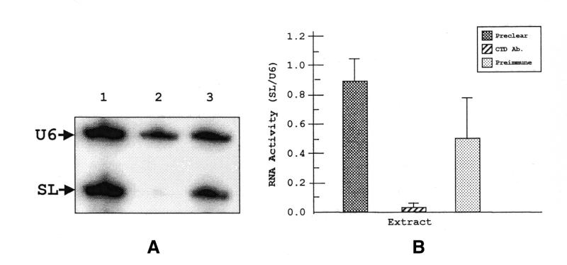
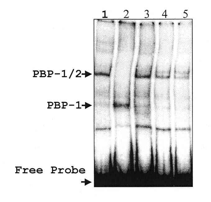
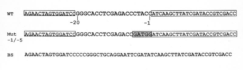
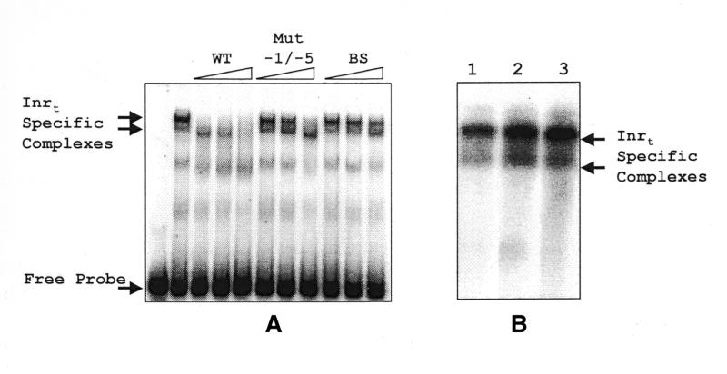
Typical general transcription factors, such as TATA binding protein and TFII B, have not yet been identified in any member of the Trypanosomatidae family of parasitic protozoa. Interestingly, mRNA coding genes do not appear to have discrete transcriptional start sites, although in most cases they require an RNA polymerase that has the biochemical properties of eukaryotic RNA polymerase II. A discrete transcription initiation site may not be necessary for mRNA synthesis since the sequences upstream of each transcribed coding region are trimmed from the nascent transcript when a short m(7)G-capped RNA is added during mRNA maturation. This short 39 nt m(7)G-capped RNA, the spliced leader (SL) sequence, is expressed as an approximately 100 nt long RNA from a set of reiterated, though independently transcribed, genes in the trypanosome genome. Punctuation of the 5' end of mRNAs by a m(7)G cap-containing spliced leader is a developing theme in the lower eukaryotic world; organisms as diverse as EUGLENA: and nematode worms, including Caenorhabditis elegans, utilize SL RNA in their mRNA maturation programs. Towards understanding the coordination of SL RNA and mRNA expression in trypanosomes, we have begun by characterizing SL RNA gene expression in the model trypanosome Leptomonas seymouri. Using a homologous in vitro transcription system, we demonstrate in this study that the SL RNA is transcribed by RNA polymerase II. During SL RNA transcription, accurate initiation is determined by an initiator element with a loose consensus of CYAC/AYR(+1). This element, as well as two additional basal promoter elements, is divergent in sequence from the basal transcription elements seen in other eukaryotic gene promoters. We show here that the in vitro transcription extract contains a binding activity that is specific for the initiator element and thus may participate in recruiting RNA polymerase II to the SL RNA gene promoter.


Figures

Figure 1
Figure 1
Identification of the L.seymouri RNA pol II largest subunit gene. SalI-digested recombinant λ phage 7-1-2 was size-separated on a 0.8% agarose gel and stained with SYBR Green (FMC Bioproducts, Chicago, IL) (lanes 1 and 2). The 4.3 kb phage DNA fragment in lane 2 is indicated. Lane 3 is the hybridization result when the DNA in lane 2 was transferred to Nytran and probed with radiolabeled pSKPIIa1. Lane 4 contains SalI-digested L.seymouri genomic DNA also probed with radiolabeled oligonucleotide SKPIIa1. The genomic DNA-hybridized fragment is indicated. Markers are λ phage DNA digested with HindIII.
Figure 2
Figure 2
Protein sequence comparison of the L.seymouri RNA pol II subunit CTD and other trypanosomatid species. T.brucei (18), Leishmania major (67) and Crithidia fasiculata (68) CTD sequences were compared with L.seymouri. Leptomonas seymouri amino acid numbers, on the right, are based on the entire RNA pol II largest subunit protein sequence. Amino acid identity with L.seymouri is represented by a dash. Gaps in sequences (represented by dots) were inserted to improve alignment. The di-serine motifs are underlined. The largest region of identity among the trypanosomatids is boxed. Sequences were aligned using the Gapped BLAST algorithm.
Figure 3
Figure 3
Purification of HisCTD using a nickel column and western analysis of recombinant protein. (A) Bacterial extract and nickel column eluates were electrophoresed on a 10% SDS–polyacrylamide gel, fixed in 10% acetic acid/10% methanol and stained with Coomassie blue. Samples are as follows: bacterial extract (lane 1), flow-through fraction (lane 2), wash fraction (lane 3), 1 M imidazole eluate (lane 4). The recombinant HisCTD protein band is indicated. (B) Recombinant HisCTD was electrophoresed as in (A), transferred to PVDF membrane and probed with either pre-immune serum (left) or anti-CTD antibody (right). The recombinant protein band is indicated. (C) Nuclear extract was electrophoresed on a 7% SDS–polyacrylamide gel transferred and probed with either the pre-immune serum (right) or anti-CTD antibody (left). Lanes 1 and 2 contain two independent extracts. (D) The blot in (C) was stripped and reprobed with anti-CTD antibody that had been blocked with either the HisORF 50 protein (lane 1) or the HisCTD protein (lane 2).
Figure 4
Figure 4
Western blot analysis of immunoprecipitated (IP) extracts. IP complexes and supernatants were electrophoresed on a 7% SDS–polyacrylamide gel, transferred overnight and probed with anti-CTD antibody (10 weeks p.i.). The extracts used were as follows: nuclear extract (lane 1), pre-cleared extract (lane 2), anti-CTD IP supernatant (lane 3), pre-immune IP supernatant (lane 4), anti-CTD IP pellet (lane 5), pre-immune IP pellet (lane 6). The band representing the RNA pol II largest subunit is indicated.
Figure 5
Figure 5
In vitro transcription with immunodepleted extracts. (A) In vitro transcription products were primer extended with a 32P-labeled primer (VB207gi) specific to a 19 nt tag present in both SL and U6 snRNA genes. The extension products were electrophoresed on a 10% polyacrylamide–7 M urea denaturing gel and exposed to a phosphorimager screen. Specific SL RNA and U6 RNA primer extended products are indicated. The extract used in each reaction was: preclear (lane 1), anti-CTD depleted (lane 2) and pre-immune depleted (lane 3). (B) Quantitation of the primer extended products. RNA activity is represented as arbitrary units of phosphorescence in a ratio of SL RNA activity divided by U6 snRNA activity. The P-values with respect to the pre-clear are 0.016 and 0.18 for anti-CTD antibody and pre-immune treated extracts, respectively.
Figure 6
Figure 6
PBP-1 and PBP-2 EMSAs. IP extracts were used in an EMSA reaction along with partially purified PBP-1 and PBP-1/-2. Extracts were incubated with a 95 bp 32P-labeled probe containing the promoter elements, PBP-1 and PBP-2, and electrophoresed on a native 4% polyacrylamide gel. The reactions contain the following proteins: affinity purified PBP-1/-2 (lane 1), PBP-1 purified from a non-specific double-stranded DNA column (lane 2), pre-cleared extract (lane 3), anti-CTD IP supernatant (lane 4), pre-immune IP supernatant (lane 5).
Figure 7
Figure 7
Diagram of DNA competitor fragments used in EMSAs. The wild-type (WT) DNA sequence used in Inrt competitions is indicated in the first line of the figure. SL RNA promoter sequences from –1 to –20 bp are flanked by sequences from the pBlueScript II SK+ (indicated by boxes). The names of the competitor fragments are indicated to the left of the sequences. The mutated sequences in Mut –1/–5 are indicated by the shaded region.
Figure 8
Figure 8
EMSA identifies Inrt binding activities within Leptomonas nuclear extracts. (A) Nuclear extracts were incubated with an Inrt-containing probe (as described in Materials and Methods) alone or in the presence of DNA competitors. The competitor amounts used in competitions were 5, 10 and 20 ng. The competitors used were: WT, the wild-type sequence; Mut –1/–5, which contains mutated sequences in the –1 to –5 bp region of the Inrt; and BS, BlueScript sequence. (B) Nuclear extract alone (lane 1), depleted with anti-CTD antibody (lane 2) or depleted with pre-immune serum (lane 3) was incubated with the Inrt-containing probe used in (A). The arrows indicate the Inrt-specific complexes.
Figure 9
Figure 9
Comparison of the α-amanitin resistance coding region of the RNA pol II largest subunit. Mouse (Mus musculus) (35) and Trichomonas (T.vaginalis) (46) sequences from domain E of the RNA pol II largest subunit are compared with L.seymouri amino acid sequences 685–760. Boxed residues indicate amino acid residues found to be individually mutated in four independently isolated α-amanitin-resistant mouse mutants (38). The sequences were aligned using the Gapped BLAST algorithm. Gaps in sequences (represented by dots) were inserted to improve alignment. Dashes represent identity with L.seymouri sequences.

References

    1. Vanhamme L. and Pays,E. (1995) Control of gene expression in trypanosomes. Microbiol. Rev., 59, 223–240. - PMC - PubMed
    1. Bangs J.D., Crain,P.F., Hashizume,T., McCloskey,J.A. and Boothroyd,J.C. (1992) Mass spectrometry of mRNA cap 4 from trypanosomatids reveals two novel nucleosides. J. Biol. Chem., 267, 9805–9815. - PubMed
    1. Günzl A., Ullu,E., Dörner,M., Fragoso,S., Hoffmann,K., Milner,J., Morita,Y., Nguu,E., Vanacova,S., Wünsch,S., Dare,A., Kwon,H. and Tschudi,C. (1997) Transcription of the Trypanosoma brucei spliced leader RNA gene is dependent only on the presence of upstream regulatory elements. Mol. Biochem. Parasitol., 85, 67–76. - PubMed
    1. Saito R.M., Elgort,M.G. and Campbell,D.A. (1994) A conserved upstream element is essential for transcription of the Leishmania tarentolae mini-exon. EMBO J., 13, 5460–5469. - PMC - PubMed
    1. Agami R., Aly,R., Halman,S. and Shapira,M. (1994) Functional analysis of cis-acting DNA elements required for expression of the SL RNA gene in the parasitic protozoan Leishmania amazonensis. Nucleic Acids Res., 22, 1959–1965. - PMC - PubMed

Publication types

MeSH terms

Associated data