. 2001 Apr 1;29(7):E35.
doi: 10.1093/nar/29.7.e35.

The length of telomeric G-rich strand 3'-overhang measured by oligonucleotide ligation assay

Affiliations

The length of telomeric G-rich strand 3'-overhang measured by oligonucleotide ligation assay

G Cimino-Reale et al. Nucleic Acids Res. .

Abstract

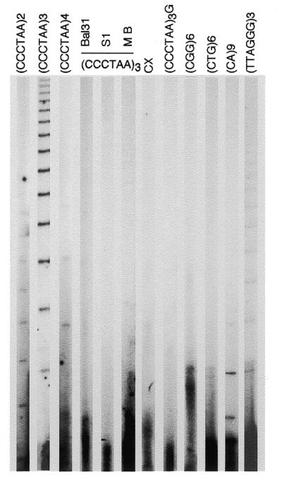
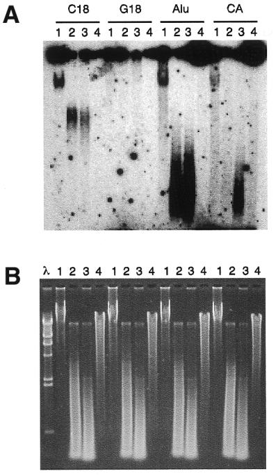
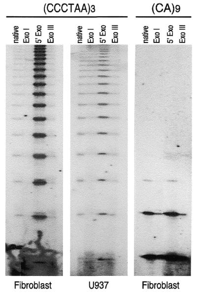
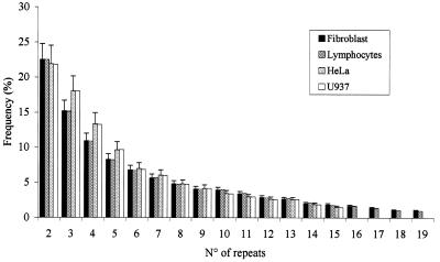
A typical G-rich telomeric DNA strand, which runs 5'-->3' toward the chromosome ends, protrudes by several nucleotides in lower eukaryotes. In human chromosomes long G-rich 3'-overhangs have been found. Apart from the standard G-rich tail, several non-canonical terminal structures have been proposed. However, the mechanism of long-tail formation, the presence and the role of these structures in telomere maintenance or shortening are not completely understood. In a search for a simple method to accurately measure the 3'-overhang we have established a protocol based on the ligation of telomeric oligonucleotide hybridized to non-denatured DNA under stringent conditions (oligonucleotide ligation assay with telomeric repeat oligonucleotide). This method enabled us to detect a large proportion of G-rich single-stranded telomeric DNA that was as short as 24 nt. Nevertheless, we showed G-tails longer than 400 nt. In all tested cells the lengths ranging from 108 to 270 nt represented only 37% of the whole molecule population, while 56-62% were <90 nt. Our protocol provides a simple and sensitive method for measuring the length of naturally occurring unpaired repeated DNA.


Figures

Figure 1
Figure 1
Oligonucleotide ligation assay to non-denaturated DNA. Ladders of ligated oligonucleotide were obtained by running the samples on 6% denaturating polyacrylamide gel. An aliquot of 5 µg of undenaturated DNA from peripheral blood lymphocytes was hybridized with 32P end-labeled oligonucleotide. OLA was performed as described in the text. Oligonucleotide probes consisting of two, three or four CCCTAA telomeric repeats were used as indicated. DNA pre-treated with Bal31, S1 and Mung Bean (MB) Exonucleases was used for OLA to the (CCCTAA)3 probe. Other probes were: CCCTTACCCTTACCCTTACCCTAA (CX), (CCCTAA)3G,(CGG)6, (CTG)6, (CA)9 and (TTAGGG)3. Each lane was loaded with 2 µl of formamide containing samples representing 20% of the OLA reaction. For autoradiography, gels were exposed for 5 days at –70°C to Biomax film and a Biomax intensifying screen.
Figure 2
Figure 2
Non-denaturing solution hybridization analysis. Solution hybridization of non-denaturating fibroblast DNA is shown. All DNA samples were hybridized to 32P-labeled probes [C18 for (CCCTAA)3, G18 for (TTAGGG)3, Alu for CGACCTCGAGATCCYRGCTCACTGCAA and CA for (CA)9]. After overnight hybridization at 50°C, the samples were separated by electrophoresis in 1% agarose and detected by autoradiography. (A) For each probe, lanes 1 show reactions with undigested DNA. Hybridizations to digested DNA with DpnII (lanes 2), with AluI (lanes 3) and with Bal31 Exonuclease (lanes 4) are shown. In all cases, 5 µg of DNA was used. (B) Ethidium bromide staining of the gel shown in (A). The λ–HindIII molecular marker is also shown.
Figure 3
Figure 3
Effects of enzymatic treatments of DNA on OLA. Treatments were performed as reported in the text. In all reactions, 5 µg of treated DNA were analyzed by OLA, either probing with (CCCTAA)3, or with (CA)9 oligonucleotide. Since no difference has been observed between fibroblasts and lymphocytes or between HeLa and U937 cells probing for telomeric repeats, and for all kinds of cell probing for CA repeats, only one kind of cell type for each category is shown. The ladders of ligated products obtained by autoradiography of denaturing gels are shown. Native, undigested DNA; ExoI: Exonuclease I; 5′ Exo: T7 (Gene6) Exonuclease; Exo III: Exonuclease III.
Figure 4
Figure 4
Size distribution of G-rich 3′ overhangs in four human cell types. The percentages of G-rich tails of different lengths are reported for fibroblasts, lymphocytes, HeLa and U937 cells. The frequency of overhangs of different length is obtained by normalizing the intensity of each single band to the sum of intensity of all the bands of the ladder. On the x-axis the number of oligonucleotides ligated by the OLA are reported.

References

    1. Muller H.J. (1938) The remaking of chromosomes. The Collecting Net, Woods Hope (USA), 13, 181–195.
    1. McClintock B. (1939) The behavior in successive nuclear divisions of a chromosome broken at meiosis. Proc. Natl Acad. Sci. USA, 25, 405–416. - PMC - PubMed
    1. Blackburn E.H. (1991) Structure and function of telomeres. Nature, 350, 569–573. - PubMed
    1. Blackburn E.H. (1994) Telomeres: no end in sight. Cell, 77, 621–623. - PubMed
    1. Zakian V.A. (1995) Telomeres: beginning to understand the end. Science, 270, 1601–1607. - PubMed

Publication types