. 2001 Jan;10(1):108-15.
doi: 10.1110/ps.37301.

Structure of soybean seed coat peroxidase: a plant peroxidase with unusual stability and haem-apoprotein interactions

Affiliations

Structure of soybean seed coat peroxidase: a plant peroxidase with unusual stability and haem-apoprotein interactions

A Henriksen et al. Protein Sci. 2001 Jan.

Abstract

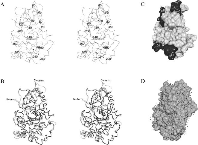
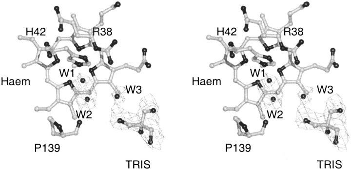
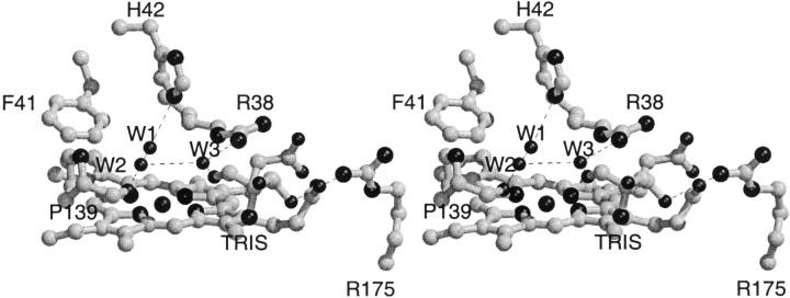
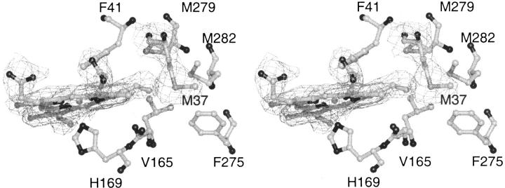
Soybean seed coat peroxidase (SBP) is a peroxidase with extraordinary stability and catalytic properties. It belongs to the family of class III plant peroxidases that can oxidize a wide variety of organic and inorganic substrates using hydrogen peroxide. Because the plant enzyme is a heterogeneous glycoprotein, SBP was produced recombinant in Escherichia coli for the present crystallographic study. The three-dimensional structure of SBP shows a bound tris(hydroxymethyl)aminomethane molecule (TRIS). This TRIS molecule has hydrogen bonds to active site residues corresponding to the residues that interact with the small phenolic substrate ferulic acid in the horseradish peroxidase C (HRPC):ferulic acid complex. TRIS is positioned in what has been described as a secondary substrate-binding site in HRPC, and the structure of the SBP:TRIS complex indicates that this secondary substrate-binding site could be of functional importance. SBP has one of the most solvent accessible delta-meso haem edge (the site of electron transfer from reducing substrates to the enzymatic intermediates compound I and II) so far described for a plant peroxidase and structural alignment suggests that the volume of Ile74 is a factor that influences the solvent accessibility of this important site. A contact between haem C8 vinyl and the sulphur atom of Met37 is observed in the SBP structure. This interaction might affect the stability of the haem group by stabilisation/delocalisation of the porphyrin pi-cation of compound I.


Figures

Fig. 1.
Fig. 1.
(A) A stereo drawing of the Cα trace of SBP. The structure is viewed facing the substrate access channel. Residue numbers are included for every 20th amino acid. (B) Stereo drawing of SBP (light grey) with HRPC (black) superimposed. The part of the HRPC trace that corresponds to variable regions defined by a STAMP alignment (Russell and Barton 1992) of class III peroxidases is colored dark grey. Conserved secondary structural elements are labeled according to the labeling of secondary structural elements in peanut peroxidase. R38, H42 and haem are shown in ball and stick like the two structural Ca2 + that appear as black spheres. (C) The solvent accessible surface of SBP in the same orientation as A and B. The surface of variable regions is black. (D) The solvent-accessible surface of HRPC with ferulic acid bound in its substrate-binding site as defined by the HRPC-FA1 binding mode (Henriksen et al. 1999). This structure is rotated slightly compared to the orientations of A, B, and C to give a better look into the substrate binding site. The small black spheres are water molecules found in the HRPC-FA structure. A and B were prepared with MOLSCRIPT (Kraulis 1991) and Raster3D (Merritt and Murphy 1994). C and D were prepared with the program DINO (Philippsen 1999).
Fig. 2.
Fig. 2.
The 1σ 2Fo-Fc electron density in the active site cavity of SBP. The two water molecules closest to the haem iron are not resolved properly and probably are corresponding to a single disordered water molecule.
Fig. 3.
Fig. 3.
Stereo drawing of the hydrogen bonding pattern in the active site of SBP.
Fig. 4.
Fig. 4.
Stereo drawing of the superposition of the active sites of SBP (light grey) and HRPC:FA2 complex (dark grey). The TRIS molecule that is found in the SBP structure is hydrogen bonded to the active site arginine through a water molecule. H42 is rotated 45° about χ2 in SBP relative to HRPC and as result, F41 is displaced ∼0.5 Å. The substitution of A74 in HRPC with an isoleucine in SBP is likely to give rise to the H42 rotation. The overall result is a more solvent, exposed δ-meso haem edge in SBP.
Fig. 5.
Fig. 5.
Resonance Raman spectra at room temperature of SBP in 0.1 M bicine buffer pH 7.0, 1 mM CaCl2 (a), and of a single crystal (b). Experimental conditions: 5 cm-1 spectral resolution (a) 514.5 nm excitation: 70 mW laser power at the sample, 45 s/0.5 cm-1 accumulation time; (b) 50 μW laser power on the crystal surface, 1560 s accumulation time.
Fig. 6.
Fig. 6.
Stereo picture of the 1σ 2Fo-Fc electron density surrounding the haem group and M37 in SBP. M37 has a closest distance of 3.9 Å to the C8 haem vinyl substituent.

References

    1. Ator, M.A. and Ortiz de Montellano, P.R. 1987. Protein control of prosthetic heme reactivity. Reaction of substrates with the heme edge of horseradish peroxidase. J. Biol. Chem. 262 1542–1551. - PubMed
    1. Barton, G.J. 1993. ALSCRIPT. A tool to format multiple sequence alignments. Protein Engineering 6 37–40. - PubMed
    1. Bernards, M.A., Fleming, W.D., Llewellyn, D.B., Priefer, R., Yang, X., Sabatino, A., and Plourde, G.L. 1999. Biochemical characterization of the suberization-associated anionic peroxidase of potato. Plant Physiol. 121 135–146. - PMC - PubMed
    1. Brünger, A.T. 1992. X-PLOR Version 3.1. A system for x-ray crystallography and NMR, Yale University Press, New Haven, CT.
    1. Brünger, A.T., Adams, P.D., Clore, G.M., DeLano, W.L., Gros, P., Grosse-Kunstleve, R.W., Jiang, J.-S., Kuszewski, J., Nilges, M., Pannu, N.S., Read, R.J., Rice, L.M., Simonson, T., and Warren, G.L. 1998. Crystallography & NMR system: A new software suite for macromolecular structure determination. Acta Crystallogr. D54 905–921. - PubMed

Publication types

Associated data