. 2001 Feb;10(2):262-76.
doi: 10.1110/ps.35801.

Plasticity of quaternary structure: twenty-two ways to form a LacI dimer

Affiliations

Plasticity of quaternary structure: twenty-two ways to form a LacI dimer

L Swint-Kruse et al. Protein Sci. 2001 Feb.

Abstract

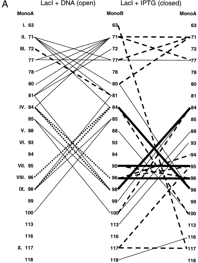
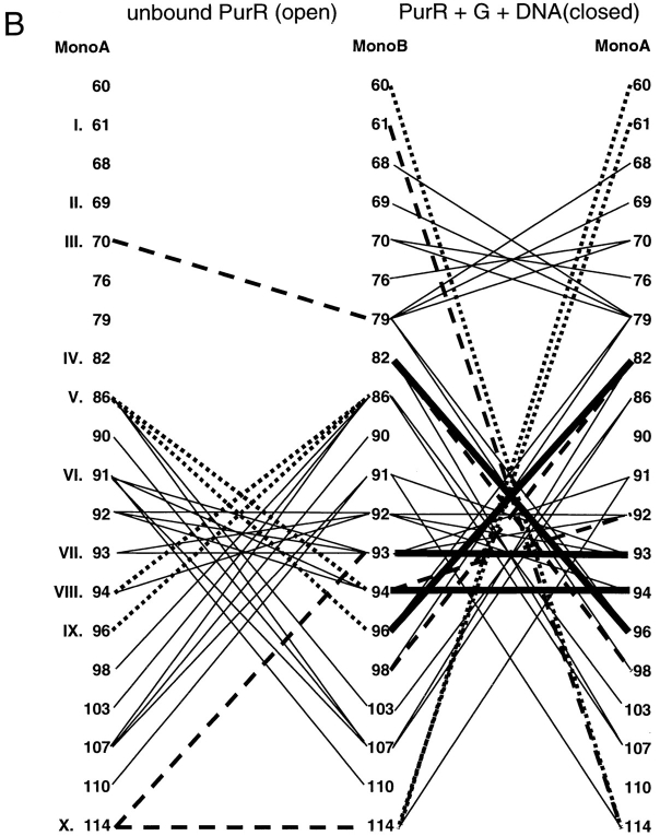
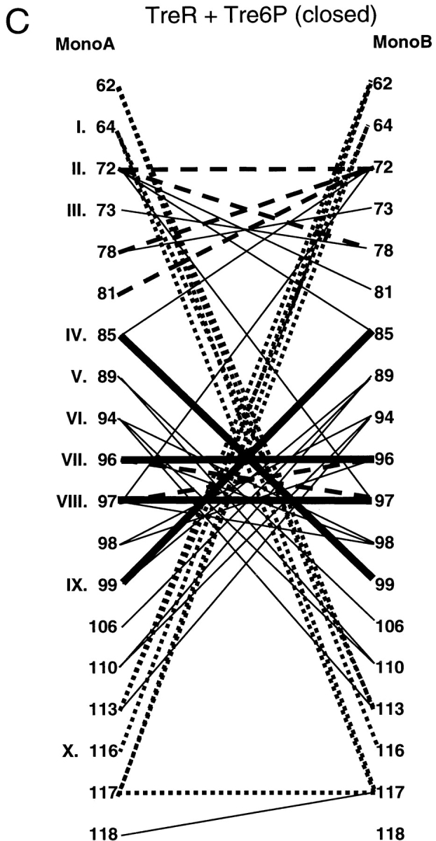
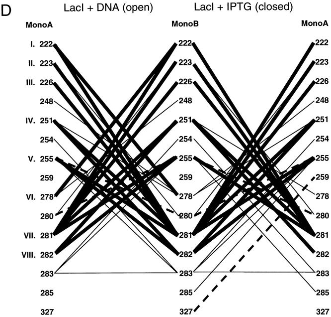
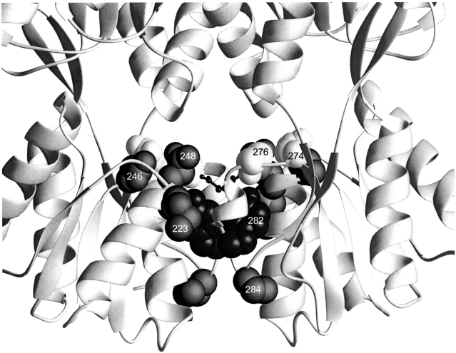
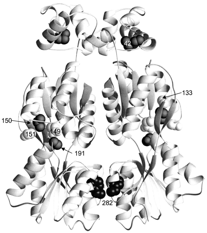
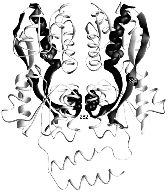
The repressor proteins of the LacI/GalR family exhibit significant similarity in their secondary and tertiary structures despite less than 35% identity in their primary sequences. Furthermore, the core domains of these oligomeric repressors, which mediate dimerization, are homologous with the monomeric periplasmic binding proteins, extending the issue of plasticity to quaternary structure. To elucidate the determinants of assembly, a structure-based alignment has been created for three repressors and four periplasmic binding proteins. Contact maps have also been constructed for the three repressor interfaces to distinguish any conserved interactions. These analyses show few strict requirements for assembly of the core N-subdomain interface. The interfaces of repressor core C-subdomains are well conserved at the structural level, and their primary sequences differ significantly from the monomeric periplasmic binding proteins at positions equivalent to LacI 281 and 282. However, previous biochemical and phenotypic analyses indicate that LacI tolerates many mutations at 281. Mutations at LacI 282 were shown to abrogate assembly, but for Y282D this could be compensated by a second-site mutation in the core N-subdomain at K84 to L or A. Using the link between LacI assembly and function, we have further identified 22 second-site mutations that compensate the Y282D dimerization defect in vivo. The sites of these mutations fall into several structural regions, each of which may influence assembly by a different mechanism. Thus, the 360-amino acid scaffold of LacI allows plasticity of its quaternary structure. The periplasmic binding proteins may require only minimal changes to facilitate oligomerization similar to the repressor proteins.


Figures

Fig. 1.
Fig. 1.
Structure of LacI. The LacI homotetramer is best described as a dimer of dimers (Friedman et al. 1995; Lewis et al. 1996). Monomers of the right dimer are black and gray to show how the N-terminal DNA-binding domains "cross over" the core domains of their partners. DNA is indicated at the top of the figure with balls and sticks. Each monomer comprises 360 amino acids. The first 60 amino acids are involved in DNA binding (Geisler and Weber 1977; Jovin et al. 1977; Ogata and Gilbert 1978; Khoury et al. 1991); a complete DNA binding site is composed of the two DNA-binding domains of a dimer (dark gray on left dimer). Residues 61–340 make up the core domain (light gray on left dimer; Platt et al. 1973; Files and Weber 1976), which contains the inducer binding site (one per monomer) and the monomer–monomer interface. The two inducer binding sites of the left dimer are indicated (*). The remaining residues (341–360) encompass an LHR sequence that forms the tetramerization domain (bottom of figure; Alberti et al. 1991, 1993; Chakerian et al. 1991; Chen et al. 1992a). This structure was generated from the pdb file 1lbg (Lewis et al. 1996).
Fig. 2.
Fig. 2.
Mutations in the monomer–monomer interface of LacI. This structure depicts one of the two dimers of the IPTG-bound structure of LacI (1lbh; Lewis et al. 1996). The N-terminal DNA binding domain is not resolved for this structure. Mutations affecting interface strength and assembly (at positions 84, 98, 74, 282, and 354) are indicated with spheres. D278, which forms an ion pair with H74, is indicated with the smaller balls and sticks.
Fig. 3.
Fig. 3.
Structure-based alignment of repressor and periplasmic binding proteins. Structure-based alignments were made by using the Dali server at the EMBL (http://www.emblheidelberg.de/Services/index.html) between the liganded "closed" structure of LacI (1lbh; Lewis et al. 1996) and the following proteins: PurR (1wet; Schumacher et al. 1994), TreR (1byk; Hars et al. 1998), RBP (2dri; Mowbray and Cole 1992), ABP (1abe; Newcomer et al. 1981; Quiocho and Vyas 1984), GBP (1glg; Vyas et al. 1988), and ALBP (1rpj; Chaudhuri et al. 1999). Gaps in the sequence of one protein relative to another are indicated with dashes. Dots are used to indicate loops "inserted" on ABP (41–66 and 239–249) that are too long to include on this figure or to indicate where sequence is not shown for simplicity (e.g., LacI 286–325). For the repressor proteins, residues with side chain or backbone contacts within 3.5 Å or hydrophobic contacts within 4.5 Å of the partner monomer are noted. Interactions of the liganded (closed) structure are represented with black text on a gray box; those of the unliganded (open) structure are indicated with white text on a gray box; and those present in both structures are shown as white text on a black box. The open structures used for LacI and PurR are 1efa and 1dbq, respectively (Schumacher et al. 1995; Bell and Lewis 2000). An open structure is not available for TreR. Two structures of closed, inducer-bound LacI are available, 1tlf and 1lbh (Friedman et al. 1995; Lewis et al. 1996). Contacts from both structures are included on this map. (A) Core-to-core contacts in the core N- and C-subdomains. Sites involved in the interfaces of all three repressors are indicated with Roman numerals I–X for the N-subdomain and I–VIII for the C-subdomain. (B) Monomer–monomer contacts between the HTH and core domains for LacI (open) and PurR (closed) are indicated, as well as LacI (closed) contacts between the core C-subdomain and the LHR. The latter are not available for the DNA-bound (open) structure of the protein.
Fig. 3.
Fig. 3.
Structure-based alignment of repressor and periplasmic binding proteins. Structure-based alignments were made by using the Dali server at the EMBL (http://www.emblheidelberg.de/Services/index.html) between the liganded "closed" structure of LacI (1lbh; Lewis et al. 1996) and the following proteins: PurR (1wet; Schumacher et al. 1994), TreR (1byk; Hars et al. 1998), RBP (2dri; Mowbray and Cole 1992), ABP (1abe; Newcomer et al. 1981; Quiocho and Vyas 1984), GBP (1glg; Vyas et al. 1988), and ALBP (1rpj; Chaudhuri et al. 1999). Gaps in the sequence of one protein relative to another are indicated with dashes. Dots are used to indicate loops "inserted" on ABP (41–66 and 239–249) that are too long to include on this figure or to indicate where sequence is not shown for simplicity (e.g., LacI 286–325). For the repressor proteins, residues with side chain or backbone contacts within 3.5 Å or hydrophobic contacts within 4.5 Å of the partner monomer are noted. Interactions of the liganded (closed) structure are represented with black text on a gray box; those of the unliganded (open) structure are indicated with white text on a gray box; and those present in both structures are shown as white text on a black box. The open structures used for LacI and PurR are 1efa and 1dbq, respectively (Schumacher et al. 1995; Bell and Lewis 2000). An open structure is not available for TreR. Two structures of closed, inducer-bound LacI are available, 1tlf and 1lbh (Friedman et al. 1995; Lewis et al. 1996). Contacts from both structures are included on this map. (A) Core-to-core contacts in the core N- and C-subdomains. Sites involved in the interfaces of all three repressors are indicated with Roman numerals I–X for the N-subdomain and I–VIII for the C-subdomain. (B) Monomer–monomer contacts between the HTH and core domains for LacI (open) and PurR (closed) are indicated, as well as LacI (closed) contacts between the core C-subdomain and the LHR. The latter are not available for the DNA-bound (open) structure of the protein.
Fig. 4.
Fig. 4.
Interface contact maps. Specific interactions of the three repressors are mapped for their core N- and C-interfaces. Proteins and structures used are the same as those used in Figure 3 ▶. Amino acids common to the interfaces of all three proteins are indicated with Roman numerals that correspond to Figure 3 ▶. For LacI and PurR, the left side of the diagram represents the interface of the unliganded open structure, and the right side of the diagram is of the liganded, closed structure. Thick, solid lines represent interactions that are conserved between all three repressors. Dashed lines indicate interactions in common between any two repressors, and the dotted lines depict interactions that have significant similarities between any two proteins. Interactions unique to one repressor are symbolized with thin black lines. These contact maps do not include the cross-subdomain interactions between LacI 74 and 278 and PurR 70 and 278.
Fig. 4.
Fig. 4.
Interface contact maps. Specific interactions of the three repressors are mapped for their core N- and C-interfaces. Proteins and structures used are the same as those used in Figure 3 ▶. Amino acids common to the interfaces of all three proteins are indicated with Roman numerals that correspond to Figure 3 ▶. For LacI and PurR, the left side of the diagram represents the interface of the unliganded open structure, and the right side of the diagram is of the liganded, closed structure. Thick, solid lines represent interactions that are conserved between all three repressors. Dashed lines indicate interactions in common between any two repressors, and the dotted lines depict interactions that have significant similarities between any two proteins. Interactions unique to one repressor are symbolized with thin black lines. These contact maps do not include the cross-subdomain interactions between LacI 74 and 278 and PurR 70 and 278.
Fig. 5.
Fig. 5.
Interactions of C281. In this view, the dimeric unit of LacI shown in Figure 2 ▶ is turned 90° out of the plane of the paper (1lbh; Lewis et al. 1996). The sidechains for C281 and Y282 on monomer A are indicated in gray (backbone interactions for these positions are not apparent on this figure). The rest of monomer A is depicted as a thin ribbon for clarity. Positions 222, 223, 226, 251, and 255 on monomer B are shown as black balls. Complementary interactions between B:281/282 and A:222, 223, 226, 251, and 255 are not shown.
Fig. 6.
Fig. 6.
Schematic of phenotypic screen for Y282D revertants.
Fig. 7.
Fig. 7.
Local mutants. This structure depicts the core C-subdomain interface for one of the two dimers of the DNA-bound structure of LacI (1efa; Bell and Lewis 2000). Positions of mutations that revert the phenotype of Y282D (223, 246, 248, 274, 276, and 284) are indicated with spheres. D278, which does not form an ion pair with H74 in this conformation, is indicated with the smaller balls and sticks.
Fig. 8.
Fig. 8.
Allosteric or DNA-binding mutants. This structure depicts one of the two dimers of the DNA-bound structure of LacI (1efa). The LHR that mediates tetramerization is not present in this structure, because a dimeric mutant of LacI was used (Bell and Lewis 2000). Positions of mutations that revert the phenotype of Y282D (42, 133, 149, 150, 151, and 191) are indicated with spheres.
Fig. 9.
Fig. 9.
Strands connecting core N- and C-subdomains. This structure depicts one of the two dimers of the IPTG-bound structure of LacI (1lbh; Lewis et al. 1996). The N-terminal DNA binding domain is not resolved for this structure. The three strands that connect the two subdomains of the core are indicated with thick ribbons. The positions of 296 and 321, mutations of which revert the monomeric phenotype of Y282D, are indicated with spheres on these strands.

Similar articles

Cited by

References

    1. Alberti, S., Oehler, S., von Wilcken-Bergmann, B., Krämer, H., and Müller-Hill, B. 1991. Dimer-to-tetramer assembly of Lac repressor involves a leucine heptad repeat. The New Biologist 3 57–62. - PubMed
    1. Alberti, S., Oehler, S., von Wilcken-Bergmann, B., and Müller-Hill, B. 1993. Genetic analysis of the leucine heptad repeats of Lac repressor: Evidence for a 4-helical bundle. EMBO J. 12 3227–3236. - PMC - PubMed
    1. Altschul, S.F., Gish, W., Miller, W., Myers, E.W., and Lipman, D.J. 1990. Basic local alignment search tool. J. Mol. Biol. 215 403–410. - PubMed
    1. Barbier, C.S. and Short, S.A. 1993. Characterization of cytR mutations that influence oligomerization of mutant repressor subunits. J. Bacteriol. 175 4625–4630. - PMC - PubMed
    1. Barry, J.K. and Matthews, K.S. 1999. Substitutions at histidine 74 and aspartate 278 alter ligand binding and allostery in lactose repressor protein. Biochemistry 38 3579–3590. - PubMed

Publication types

MeSH terms