. 2001 Feb;10(2):301-12.
doi: 10.1110/ps.33601.

Solvation energetics and conformational change in EF-hand proteins

Affiliations

Solvation energetics and conformational change in EF-hand proteins

A Ababou et al. Protein Sci. 2001 Feb.

Abstract

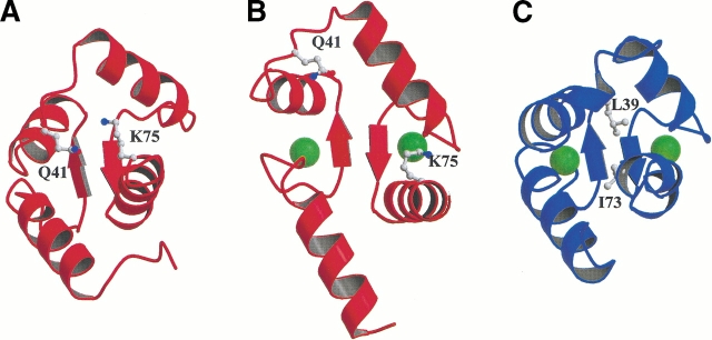
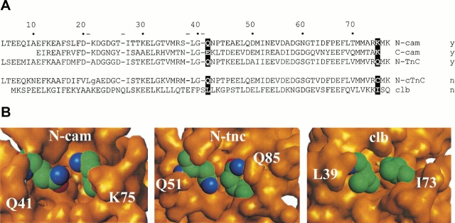
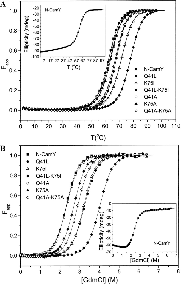
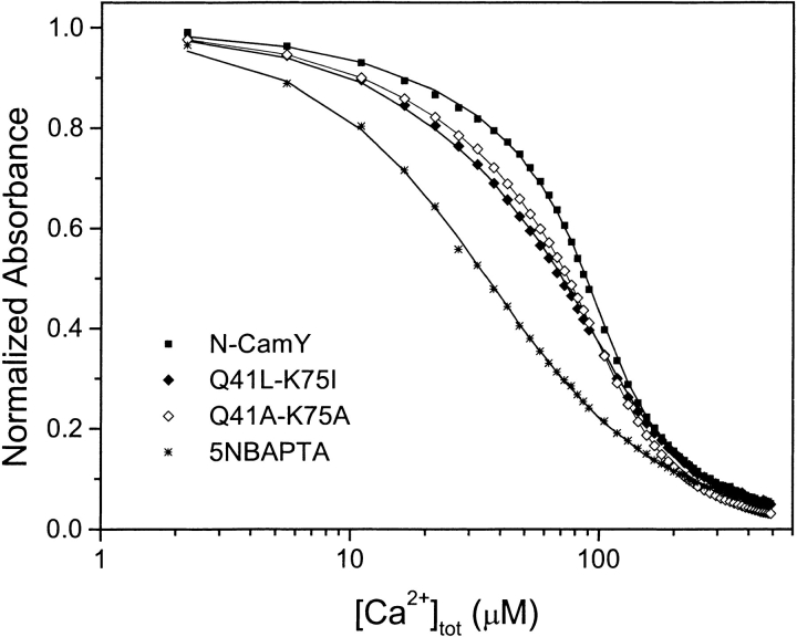
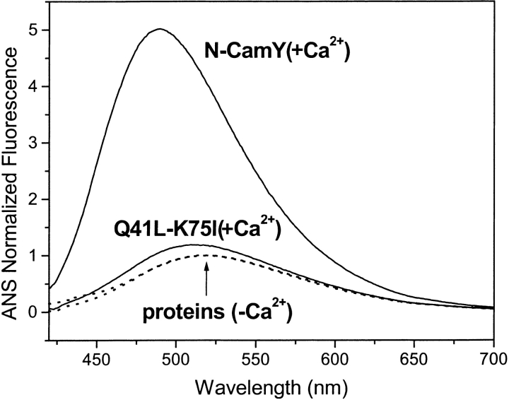
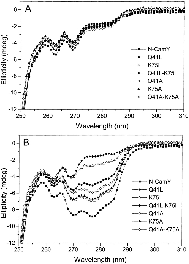
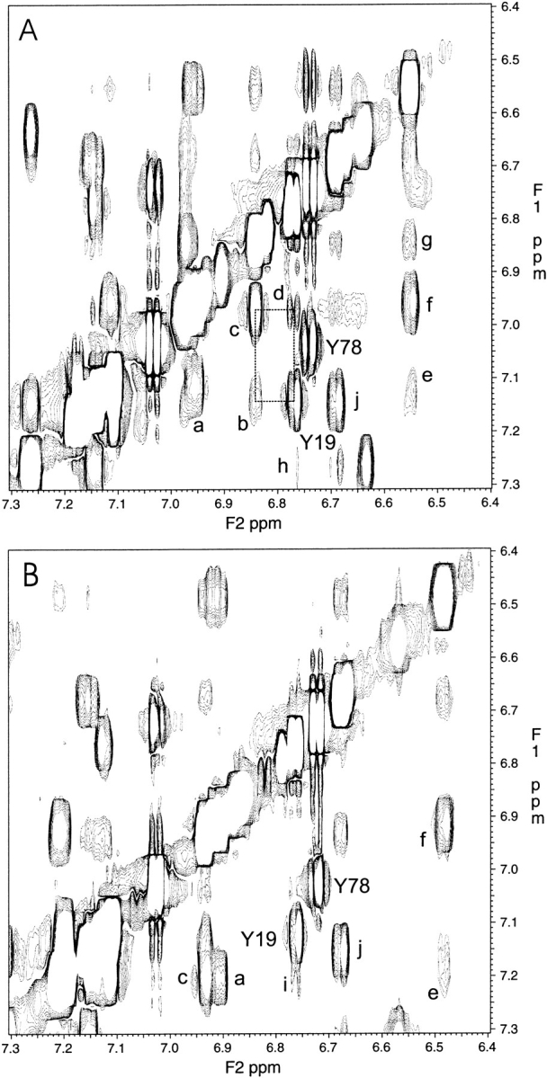
Calmodulin and other members of the EF-hand protein family are known to undergo major changes in conformation upon binding Ca(2+). However, some EF-hand proteins, such as calbindin D9k, bind Ca(2+) without a significant change in conformation. Here, we show the importance of a precise balance of solvation energetics to conformational change, using mutational analysis of partially buried polar groups in the N-terminal domain of calmodulin (N-cam). Several variants were characterized using fluorescence, circular dichroism, and NMR spectroscopy. Strikingly, the replacement of polar side chains glutamine and lysine at positions 41 and 75 with nonpolar side chains leads to dramatic enhancement of the stability of the Ca(2+)-free state, a corresponding decrease in Ca(2+)-binding affinity, and an apparent loss of ability to change conformation to the open form. The results suggest a paradigm for conformational change in which energetic strain is accumulated in one state in order to modulate the energetics of change to the alternative state.


Figures

Fig. 1.
Fig. 1.
Ca2+-induced conformational changes in the N-terminal domain of calmodulin. (A) Ribbon diagrams show the differences in the conformations of Ca2+-free N-Cam (Kuboniwa et al. 1995); (B) Ca2+-saturated N-cam (Chattopadhyaya et al. 1992); and (C) Ca2+-saturated calbindin D9k (Svensson et al. 1992). The residues Gln41 and Lys75 in N-Cam examined in this study, as well as Leu38 and Ile73 in calbindin D9k, are displayed. Green spheres represent the Ca2+ ions. The figures were made with MOLSCRIPT (Kraulis 1991) and Raster 3D (Merrit and Bacon 1997).
Fig. 2.
Fig. 2.
Partially buried polar groups in EF-hand proteins. (A) Alignment of EF-hand domain sequences: the N- and C-terminal domains of calmodulin (N-cam, C-cam), the regulatory domain of skeletal troponin C (N-tnC), the regulatory domain of cardiac troponin C (N-ctnC), and calbindin D9k (clb). The numbering scheme is based on the full-length calmodulin sequence. The positions that are the focus of this work are highlighted in black. The ability to change conformation (y) or not (n) is listed for each domain. (B) Close-ups of buried polar groups in Ca2+-free structures of N-cam and the regulatory domain of troponin C (N-tnc). Calbindin (clb), in contrast, contains nonpolar groups at structurally equivalent positions.
Fig. 3.
Fig. 3.
Position-specific changes in nonpolar surface area (ΔnpSA). (A) Change in npSA of N-cam residues, plotted as the difference in accessible surface areas calculated from the Ca2+-saturated and -free structures. (B) Predicted change in npSA for a hybrid protein clb-EF1, constructed in order to model a hypothetical open state for calbindin (see Materials and Methods). Positions 41 and 75 from N-camY are highlighted, as are their counterparts in the clb-EF1 sequence.
Fig. 4.
Fig. 4.
Stability of Ca2+-free N-camY variants. (A) Thermal Denaturation. The apparent fraction of unfolded protein Fapp for N-CamY and the variants is plotted as a function of temperature. The extent of unfolding was monitored by circular dichroism (CD) at a wavelength of 222 nm. (B) Chemical Denaturation. The Fapp as a function of GdmCl concentration is shown for each variant. The unfolding was monitored by far-UV CD at a wavelength of 232 nm. For each figure, the inset shows the uncorrected denaturation data and initial fits for the N-camY protein.
Fig. 5.
Fig. 5.
Ca2+-binding assays for representative N-camY variants. Logarithmic representations of the normalized Ca2+ competition curves of N-CamY and the mutants monitored by the absorbance of 5NBAPTA (5-nitro-BAPTA) at 430 nm. The symbols are the experimental data and the curves through the points are the least-squares fits. Binding parameters determined from the fits are listed in Table 2.
Fig. 6.
Fig. 6.
Conformational changes measured by ANS binding. Fluorescence emission spectra of ANS in the presence of protein as illustrated in the case of N-CamY and Q41L-K75I, in the absence of Ca2+ (dashed line), normalized to unity, and its relative fluorescence enhancement in the presence of Ca2+ (solid line). Table 2 lists the relative ANS intensity for all of the variants in the presence or absence of Ca2+.
Fig. 7.
Fig. 7.
Conformational changes measured by near-UV CD spectra. Spectra of all of the variants were collected with 0.1 mM EDTA (A) or 1 mM CaCl2(B).
Fig. 8.
Fig. 8.
Aromatic regions of 1H-1H NOESY spectra. Spectra are shown for Ca2+-saturated N-camY (A) and Ca2+-saturated Q41L-K75I (B). As expected from the open-state structure of N-cam, several cross-peaks between Tyr19 and other aromatics are observed (dashed box). These cross-peaks are not expected for the closed form. Assignments—based on COSY, TOCSY, and NOESY correlations, similarities with published assignments (Ikura et al. 1990, 1991; Elshorst et al. 1999), and consistency with the know structures—are as follows. For (A) Y19, Y19 (Hɛ, 6.77) to (Hδ, 7.14); Y78, Y78 (Hɛ, 6.74) to (Hδ, 7.05); a, F16 (Hɛ, 6.96) to (Hζ, 7.11); b, F68 (Hɛ, 6.84) to Y19 (Hδ, 7.14); c, F68 (Hɛ, 6.84) to (Hζ, 6.97); d, Y19 (Hɛ, 6.77) to F68 (Hζ, 6.97); e, F16 (Hδ, 6.55) to (Hζ, 7.11); f, F16 (Hδ, 6.55) to (Hɛ, 6.96); g, F16 (Hδ, 6.55) to F68 (Hɛ, 6.84); h, Y19 (Hɛ, 6.77) to F65* j, F65* (H*, 6.69) to F65* (Hɛ, 7.15). For (B) Y19, Y19 (Hɛ, 6.76) to (Hδ, 7.13); Y78, Y78 (Hɛ, 6.72) to (Hδ, 7.02); a, F16 (Hɛ, 6.92) to (Hζ, 7.21); c, F68 (H*, 6.93) to (H*, 7.21); e, F16 (Hδ, 6.48) to (Hζ, 7.21); f, F16 (Hδ, 6.48) to (Hɛ, 6.92); i, Y19 (Hɛ, 6.76) to (*); j, F65* (H*, 6.68) to F65* (Hɛ, 7.15). *Not determined or uncertainty in the assignment.
Fig. 9.
Fig. 9.
Relationship between calcium-binding and -unfolding free energies. For proteins that change conformation, a linear correlation might be expected, denoted by the solid line. However, as the closed state becomes stabilized to the extent that calcium binding does not induce a conformational change, the free energies will become uncoupled. At this point, the calcium-binding free energy will approach the intrinsic affinity of the closed state, as denoted by the dashed line.

Similar articles

Cited by

References

    1. Ackers, G.K., Doyle, M.L., Myers, D., and Daugherty, M.A. 1992. Molecular code for cooperativity in hemoglobin. Science 255 54–63. - PubMed
    1. Babu, Y.S., Bugg, C.E., and Cook, W.J. 1988. Structure of calmodulin refined at 2.2 Å resolution. J. Mol. Biol. 204 191–204. - PubMed
    1. Bayley, P., Ahlstrom, P., Martin, S.R., and Forsen, S. 1984. The kinetics of calcium binding to calmodulin: Quin 2 and ANS stopped-flow fluorescence studies. Biochem. Biophys. Res. Commun. 120: 185–91. - PubMed
    1. Biekofsky, R.R., Martin, S.R., Browne, J.P., Bayley, P.M., and Feeney, J. 1998. Ca2+ coordination to backbone carbonyl oxygen atoms in calmodulin and other EF-hand proteins: 15N chemical shifts as probes for monitoring individual-site Ca2+ coordination. Biochemistry 37 7617–29. - PubMed
    1. Blaber, M., Lindstrom, J.D., Gassner, N., Xu, J., Heinz, D.W., and Matthews, B.W. 1993. Energetic cost and structural consequences of burying a hydroxyl group within the core of a protein determined from Ala–Ser and Val–Thr substitutions in T4 lysozyme. Biochemistry 32 11363–11373. - PubMed

MeSH terms