. 2001 Jan;217(1-2):143-52.
doi: 10.1023/a:1007209321969.

Cardioprotective effects of adenosine A1 and A3 receptor activation during hypoxia in isolated rat cardiac myocytes

Affiliations

Cardioprotective effects of adenosine A1 and A3 receptor activation during hypoxia in isolated rat cardiac myocytes

N Safran et al. Mol Cell Biochem. 2001 Jan.

Abstract

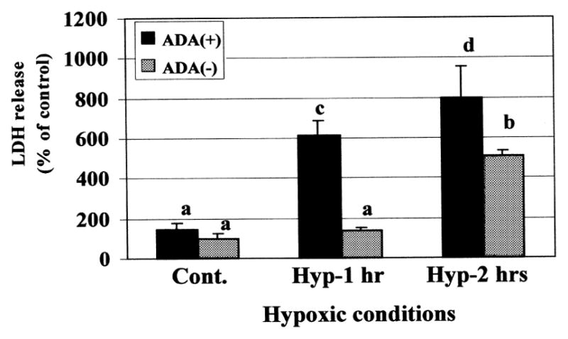
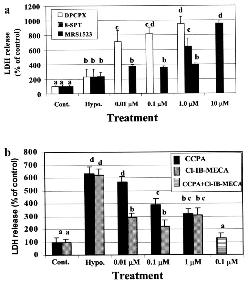
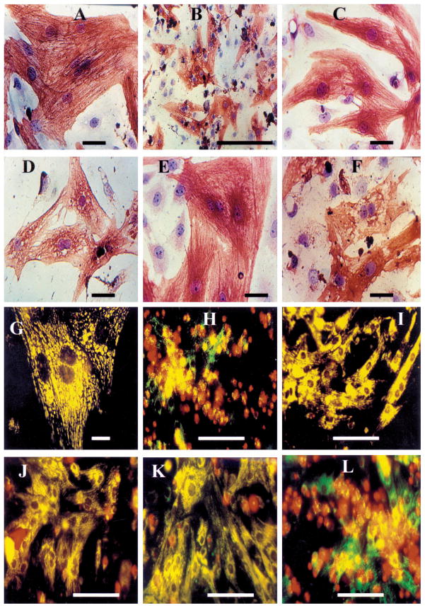
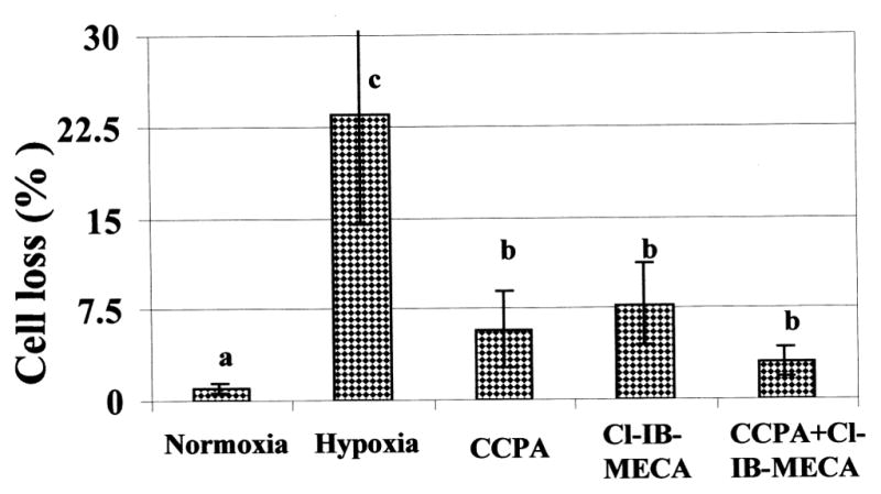
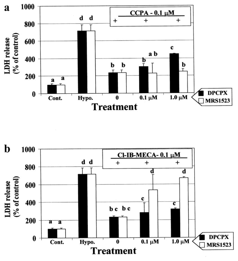
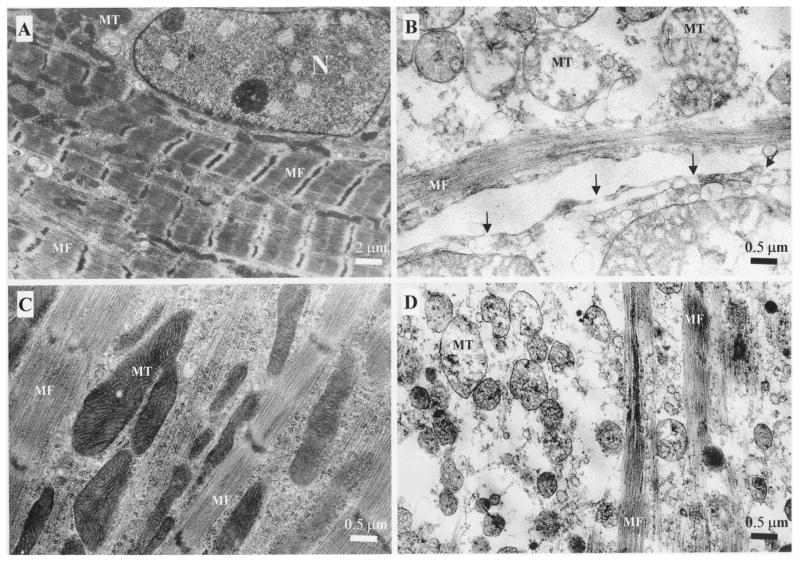
Adenosine (ADO) is a well-known regulator of a variety of physiological functions in the heart. In stress conditions, like hypoxia or ischemia, the concentration of adenosine in the extracellular fluid rises dramatically, mainly through the breakdown of ATP. The degradation of adenosine in the ischemic myocytes induced damage in these cells, but it may simultaneously exert protective effects in the heart by activation of the adenosine receptors. The contribution of ADO to stimulation of protective effects was reported in human and animal hearts, but not in rat hearts. The aim of this study was to evaluate the role of adenosine A1 and A3 receptors (A1R and A3R), in protection of isolated cardiac myocytes of newborn rats from ischemic injury. The hypoxic conditions were simulated by exposure of cultured rat cardiomyocytes (4-5 days in vitro), to an atmosphere of a N2 (95%) and CO2 (5%) mixture, in glucose-free medium for 90 min. The cardiotoxic and cardioprotective effects of ADO ligands were measured by the release of lactate dehydrogenase (LDH) into the medium. Morphological investigation includes immunohistochemistry, image analysis of living and fixed cells and electron microscopy were executed. Pretreatment with the adenosine deaminase considerably increased the hypoxic damage in the cardiomyocytes indicating the importance of extracellular adenosine. Blocking adenosine receptors with selective A1 and A3 receptor antagonists abolished the protective effects of adenosine. A1R and A3R activation during the hypoxic insult delays onset of irreversible cell injury and collapse of mitochondrial membrane potential as assessed using DASPMI fluorochrom. Cardioprotection induced by the A1R agonist, CCPA, was abolished by an A1R antagonist, DPCPX, and was not affected by an A3R antagonist, MRS 1523. Cardioprotection caused by the A3R agonist, Cl-IB-MECA, was antagonized completely by MRS 1523 and only partially by DPCPX. Activation of both A1R and A3R together was more efficient in protection against hypoxia than by each one alone. Our study indicates that activation of either A1 or A3 adenosine receptors in the rat can attenuate myocyte injury during hypoxia. Highly selective A1R and A3R agonists may have potential as cardioprotective agents against ischemia or heart surgery.


Figures

Fig. 1
Fig. 1
Duration of hypoxic conditions and the effect of adenosine deaminase (ADA) (5 units/ml) on endogenic adenosine release from cultured cardiomyocytes after prolonged exposure to hypoxic conditions. The LDH release was determined immediately after hypoxia. One hundred percent were considered as the release in the control cultures. Data are expressed as mean ± S.E.M. of at least 3 replicates in 3 separate experiments. Means with the same letter are not significantly different (p < 0.05) according to a post-hoc Tukey-Kramer test.
Fig. 2
Fig. 2
Effect of Adenosine A1R and A3R ligand in cardiac myocytes, under hypoxic conditions for 75-min in the presence of antagonist (a), and for 90-min in the presence of agonist (b). The LDH release was determined immediately after hypoxia. One hundred percent were considered as the release in the control cultures. Data are means of at least 3 replicates in 5 separate experiments. Means with the same letter are not significantly different (p < 0.05) according to a post-hoc Tukey-Kramer test.
Fig. 3
Fig. 3
Dose dependent effect of A1R (DPCPX) and A3R (MRS1523) antagonists on the cardioprotection induced by A1R (CCPA – 0.1 μM) (a) and A3R (Cl-IB-MECA – 0.1 μM) (b) agonists. The LDH release was determined after 90 min hypoxia. The 100% were determined as the release in the control cultures. Data are means of at least 3 replicates in 3 separate experiments. Means with the same letter are not significantly different (p < 0.05) according to a post-hoc Tukey-Kramer test.
Fig. 4
Fig. 4
Cardiotoxicity and cardioprotection demonstrated by morphological investigations. (A–F) Immunohistochemical staining of α-sarcomeric actin in rat cardiomyocyte cultures followed by hematoxylin counterstaining. (A) Control – normoxic conditions; (B) 90-min hypoxic conditions; (C) Presence of A1R agonist (CCPA – 0.1 μM) during hypoxic period; (D) CCPA was introduced together with A1R antagonist (DPCPX – 1 μM) during hypoxic period; (E) Presence of A3R agonist (Cl-IB-MECA – 0.1 μM) during hypoxic period; (F) Presence of A3R agonist (Cl-IB-MECA – 0.1 μM) together with A3R antagonist (MRS1523 – 1 μM) during the hypoxic period; (G–L) The accumulation of DASPMI dye in the mitochondrial matrix space (vital staining) in conjunction with propidium iodide binding to nuclei of cells whose plasma membranes have became permeable in rat cardiomyocyte cultures. (G) Control – normoxic conditions; (H) 90-min hypoxic conditions; (I) Presence of A1R agonist (CCPA – 0.1 μM) during hypoxic period; (J) CCPA was introduced in presence of A1R antagonist (DPCPX – 1 μM) during hypoxic period; (K) Presence of A3R agonist (Cl-IB-MECA – 0.1 μM) during the hypoxic period; (L) Presence of A3R agonist (Cl-IB-MECA – 0.1 μM) together with A3R antagonist (MRS1523 – 1 μM) during hypoxic period. The results were obtained in at least 3 replicates in 3 separate experiments.
Fig. 5
Fig. 5
Effect of Adenosine A1R and A3R agonists on cardiac myocytes loss after hypoxia for 90-min. A1R agonist (CCPA – 0.1 μM), or A3R agonist (Cl-IB-MECA – 0.1 μM) were introduced 10 min before and during the 90 min hypoxia. Data are means of at least 3 replicates in 3 separate experiments. Means with the same letter are not significantly different (p < 0.05) according to a post-hoc Tukey-Kramer test.
Fig. 6
Fig. 6
Electron micrographs of cardiac cells after hypoxic conditions. (A) Control cardiomyocyte in normoxic conditions; (B) Hypoxic conditions for 90-min. Gaps in the cell membrane (arrows); (C) A3R agonist (Cl-IB-MECA – 100 nM) exposure for 10-min before and during the 90-min of hypoxic conditions; (D) A3R agonist (Cl-IB-MECA – 100 nM) and A3R antagonist (MRS1523 – 1 μM) exposure for 10-min before and during the 90-min of hypoxic conditions. Nucleus (N), myofibrils (MF) and mitochondria (MT).

References

    1. Tucker AL, Robeva AS, Taylor HE, Holeton D, Bockner M, Lynch KR, Linden J. A1 adenosine receptors. Two amino acids are responsible for species differences in ligand recognition. J Biol Chem. 1994;269:27900–27906. - PubMed
    1. Berne RM. Cardiac nucleotides in hypoxia: Possible role in regulation of coronary blood flow. Am J Physiol. 1963;204:317–322. - PubMed
    1. Headrick JP. Ischemic preconditioning: Bioenergetic and metabolic changes and the role of endogenous adenosine. J Mol Cell Cardiol. 1996;28:1227–1240. - PubMed
    1. Wang J, Drake L, Sajjadi F, Firestein GS, Mullane KM, Bullough DA. Dual activation of adenosine A1 and A3 receptors mediates preconditioning of isolated cardiac myocytes. Eur J Pharmacol. 1997;320:241–248. - PubMed
    1. Tracey WR, Magee W, Masamune H, Kennedy SP, Knight DR, Buchholz RA, Hill RJ. Selective adenosine A3 receptor stimulation reduces ischemic myocardial injury in the rabbit heart. Cardiovasc Res. 1997;33:410–415. - PubMed

Publication types

MeSH terms

Substances