A comparative study of fibrous dysplasia and osteofibrous dysplasia with regard to Gsalpha mutation at the Arg201 codon: polymerase chain reaction-restriction fragment length polymorphism analysis of paraffin-embedded tissues
- PMID: 11272890
- PMCID: PMC1906902
- DOI: 10.1016/s1525-1578(10)60618-6
A comparative study of fibrous dysplasia and osteofibrous dysplasia with regard to Gsalpha mutation at the Arg201 codon: polymerase chain reaction-restriction fragment length polymorphism analysis of paraffin-embedded tissues
Abstract
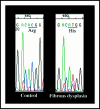
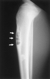
Fibrous dysplasia and osteofibrous dysplasia are both benign fibro-osseous lesions of the bone and are generally seen during childhood or adolescence. Histologically, the features of these bone lesions sometimes look quite similar, but their precise nature remains controversial. Mutation of the alpha subunit of signal-transducing G proteins (Gsalpha), with an increase in cyclic adenosine monophosphate (cAMP) formation, has been implicated in the development of multiple endocrinopathies of the Albright-McCune syndrome and in the development of fibrous dysplasia. We studied Gsalpha mutation at the Arg201. codon in seven cases of fibrous dysplasia (six monostotic lesions and one polyostotic lesion) and seven cases of osteofibrous dysplasia using formalin-fixed, paraffin-embedded tissue, by means of polymerase chain reaction-restriction fragment length polymorphism and direct sequencing analysis. All of the seven cases of fibrous dysplasia showed missense point mutations in Gsalpha at the Arg201 codon that resulted in Arg-to-His substitution in three cases and Arg-to-Cys substitution in four cases. On the other hand, the seven cases of osteofibrous dysplasia and the normal bone used as a control showed no such mutation. These data suggest that fibrous dysplasia and osteofibrous dysplasia have different pathogeneses and that the detection of Gsalpha mutation at the Arg201 codon is quite useful for distinguishing between these lesions.
Figures


References
-
- Lichtenstein L: Polyostotic fibrous dysplasia. Arch Surg 1938, 36:874-898
-
- Lichtenstein L, Jaffe HL: Fibrous dysplasia of a bone: condition affecting one, several or many bones, graver cases of which may present abnormal pigmentation of skin, premature sexual development, hyperthyroidism or still other extraskeletal abnormalities. Arch Pathol 1942, 33:777
-
- Arbright F, Bulter AM, Hampton AO, Smith P: Syndrome characterized by osteitis fibrosa disseminata, areas of pigmentation and endocrine dysfunction with precocious puberty in females. N Engl J Med 1937, 216:727-746
-
- Danon M, Crawford JD: The McCune-Albright syndrome. Ergeb inn Med Kinderheild 1987, 55:81-115 - PubMed
-
- Riminucci M, Liu B, Corsi A, Shenker A, Spiegel AM, Robey PG, Bianco P: The histopathology of fibrous dysplasia of bone in patients with activating mutations of the Gs alpha gene: site-specific patterns and recurrent histological hallmarks. J Pathol 1999, 187:249-258 - PubMed
Publication types
MeSH terms
Substances
LinkOut - more resources
Full Text Sources

