. 2001 Mar 15;15(6):652-7.
doi: 10.1101/gad.873001.

A mutation in a mitochondrial transmembrane protein is responsible for the pleiotropic hematological and skeletal phenotype of flexed-tail (f/f) mice

Affiliations

A mutation in a mitochondrial transmembrane protein is responsible for the pleiotropic hematological and skeletal phenotype of flexed-tail (f/f) mice

M D Fleming et al. Genes Dev. .

Abstract

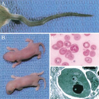
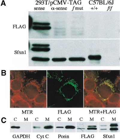
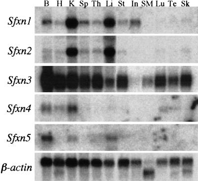
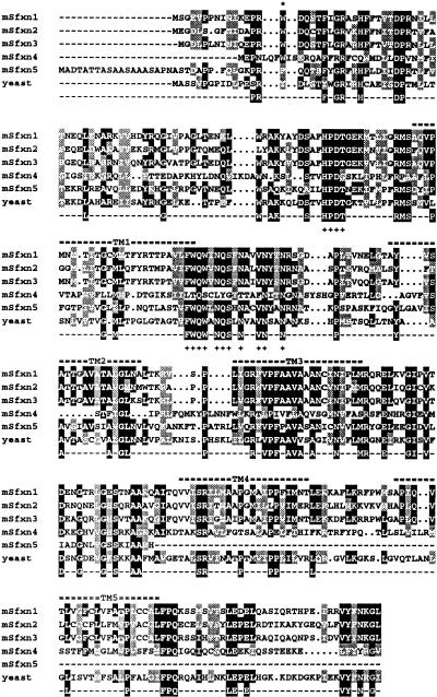
We have studied the flexed-tail (f) mouse to gain insight into mammalian mitochondrial iron metabolism. Flexed-tail animals have axial skeletal abnormalities and a transient embryonic and neonatal anemia characterized by pathologic intramitochondrial iron deposits in erythrocytes. Mitochondrial iron accumulation is the hallmark of sideroblastic anemias, which typically result from defects in heme biosynthesis or other pathways that lead to abnormal erythroid mitochondrial iron utilization. To clone the f gene, we used positional cloning techniques, and identified a frameshift mutation in a mitochondrial transmembrane protein. The mutated gene, Sfxn1, is the prototype of a novel family of evolutionarily conserved proteins present in eukaryotes.


Figures

Figure 1
Figure 1
Flexed-tail (f/f) phenotype. (A) Variable tail curvatures seen in f/f animals. (B) Pale newborn homozygous f/f animal (below) and heterozygous f/+ littermate (above). (C) Prussian blue iron-stained peripheral blood smear from a newborn f/f animal. Iron appears as dark blue granules seen in nearly all erythrocytes. (D) Transmission electron micrograph of a newborn f/f erythrocyte. A large electron-dense (black) inclusion can be seen in the mitochondrion at the bottom, and a smaller inclusion seen in the mitochondrion in the middle. This electron-dense material is typical of the appearance of iron, which was confirmed by electron energy loss spectroscopy (EELS, data not shown).
Figure 2
Figure 2
Genetic and physical maps of the flexed-tail region. (A) Genetic map of the f locus on mouse chromosome 13 determined from 1000 CAST/Ei backcross animals. PCR polymorphism markers are listed above, and recombination distances in centimorgans (cM) are below. (B) Bacterial artificial chromosome (BAC) contig spanning the f critical genetic interval. Individual sequence tagged sites (STSs) present on each BAC are indicated by black diamonds. STSs mapped in the backcross are indicated by a dashed line. (C) The intron/exon structure of the Sfxn1 gene. The gene spans approximately 35 kb and is divided into 11 exons (gray boxes). Exon 2, which was nonrecombinant with the phenotype in the backcrosses and contains the f mutation, is indicated by an asterisk. The position on the physical map of the centromeric flanking markers J2T7 and 506Hd28T7 are indicated by a single arrow and a double arrow, respectively.
Figure 3
Figure 3
Western blotting and subcellular localization of Sfxn1. (A) Western blots from HEK293T cells transfected with epitope-tagged (FLAG) wild-type Sfxn1 cloned in a sense and antisense (α-sense) orientation, as well as the f frameshift mutation (f mut), and on kidney tissue extracts from wild-type (+/+) and f/f animals on a C57BL/6J background. The FLAG-tagged protein runs as a doublet with an apparent molecular mass 4–6 kD greater than the endogenous human or mouse proteins. (B) HEK293T cells were transfected with FLAG-tagged Sfxn1 and pulsed with MitoTracker Red CMXRos (MTR). Colocalization of the epitope-tagged protein and mitochondria is seen as yellow fluorescence in the red and green merged image (MTR + FLAG). (C) Western blots of whole cell extracts (c) and purified mitochondria (m) from transfected HEK293T cells using antibodies against the cytoplasmic enzyme GAPDH, and the mitochondrial proteins porin and cytochrome C (Cyt C).
Figure 4
Figure 4
Expression of mouse sideroflexin homolog mRNAs. B, brain; H, heart; K, kidney; Sp, spleen; Th, thymus; Li, liver; St, stomach; In, intestine; SM, skeletal muscle; Lu, lung; Te, testis; Sk, skin.
Figure 5
Figure 5
Peptide alignment of mouse and yeast sideroflexin homologs. Amino acids conserved in 60% or more of the sequences are shaded black, but only those present in 80% or more of the sequences are represented in the consensus sequence. Similar amino acids present in >60% of sequences are shaded gray. Based upon its human counterpart, the mouse sideroflexin 5 sequence is a partial sequence truncated at the carboxy terminus. Predicted transmembrane domains (TM) are indicated by a double bar above the sequence alignment. The particularly well-conserved HPDT and asparagine-rich motifs are indicated by plus signs (+) under the sequences. The site of the f frameshift mutation is marked by an asterisk.

References

    1. Andrews NC. Iron homeostasis: Insights from genetics and animal models. Nat Rev (Genetics) 2000;1:208–217. - PubMed
    1. Azzi A, Glerum M, Koller R, Mertens W, Spycher S. The mitochondrial tricarboxylate carrier. J Bioenerg Biomem. 1993;25:515–524. - PubMed
    1. Bateman AE, Cole RJ. Colony forming cells in the livers of prenatal flexed (f/f) anaemic mice. Cell Tissue Kinet. 1972;5:165–173. - PubMed
    1. Bekri S, Kispal G, Lange H, Fitzsimons E, Tolmie J, Lill R, Bishop DF. Human ABC7 transporter: Gene structure and mutation causing X-linked sideroblastic anemia with ataxia with disruption of cytosolic iron–sulfur protein maturation. Blood. 2000;96:3256–3264. - PubMed
    1. Bottomley SS. Sideroblastic Anemias. In: Lee GR, editor. Wintrobe's clinical hematology. Baltimore: Williams and Wilkins; 1999. pp. 1022–1045.

Publication types

MeSH terms