. 2001 Apr;183(8):2614-23.
doi: 10.1128/JB.183.8.2614-2623.2001.

Biochemical analysis of replication factor C from the hyperthermophilic archaeon Pyrococcus furiosus

Affiliations

Biochemical analysis of replication factor C from the hyperthermophilic archaeon Pyrococcus furiosus

I K Cann et al. J Bacteriol. 2001 Apr.

Abstract

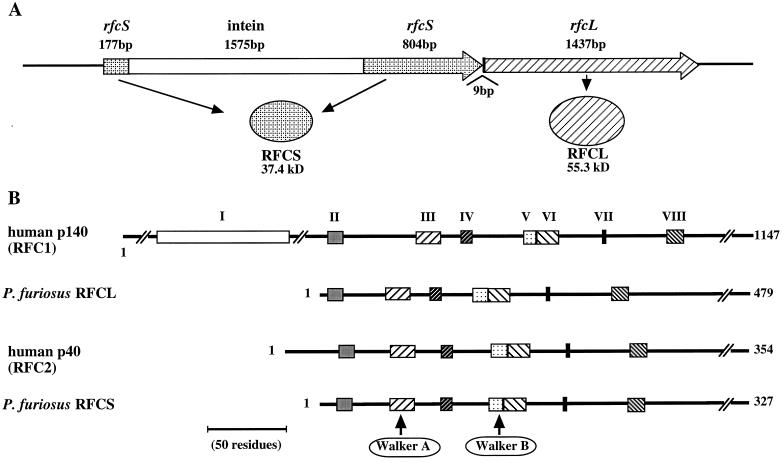
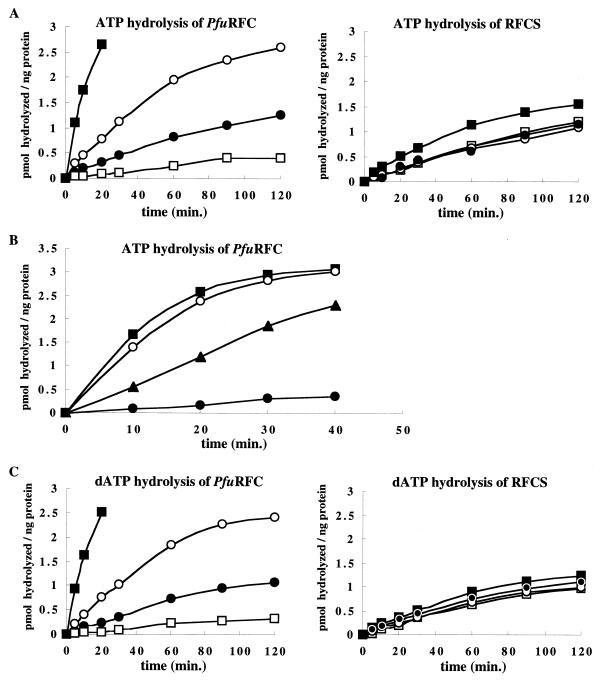
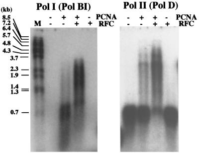
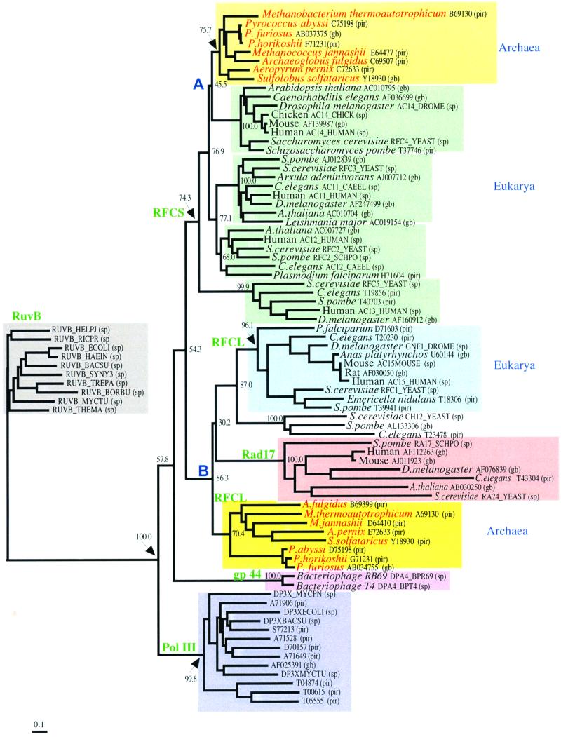
Replication factor C (RFC) and proliferating cell nuclear antigen (PCNA) are accessory proteins essential for processive DNA synthesis in the domain Eucarya. The function of RFC is to load PCNA, a processivity factor of eukaryotic DNA polymerases delta and epsilon, onto primed DNA templates. RFC-like genes, arranged in tandem in the Pyrococcus furiosus genome, were cloned and expressed individually in Escherichia coli cells to determine their roles in DNA synthesis. The P. furiosus RFC (PfuRFC) consists of a small subunit (RFCS) and a large subunit (RFCL). Highly purified RFCS possesses an ATPase activity, which was stimulated up to twofold in the presence of both single-stranded DNA (ssDNA) and P. furiosus PCNA (PfuPCNA). The ATPase activity of PfuRFC itself was as strong as that of RFCS. However, in the presence of PfuPCNA and ssDNA, PfuRFC exhibited a 10-fold increase in ATPase activity under the same conditions. RFCL formed very large complexes by itself and had an extremely weak ATPase activity, which was not stimulated by PfuPCNA and DNA. The PfuRFC stimulated PfuPCNA-dependent DNA synthesis by both polymerase I and polymerase II from P. furiosus. We propose that PfuRFC is required for efficient loading of PfuPCNA and that the role of RFC in processive DNA synthesis is conserved in Archaea and Eucarya.


Figures

FIG. 1
FIG. 1
Gene organization and amino acid sequence comparison of PfuRFC with human RFC. (A) The genes for RFCS and RFCL are arranged in tandem on the P. furiosus genome. Open reading frames are indicated by the large arrows with each encoded product. An intein gene is inserted into the site for Walker motif A of RFCS. (B) Amino acid sequences of RFCS and RFCL are compared with those of the human RFC subunits. The conserved RFC boxes are indicated by closed boxes and are numbered at the top. The amino acid lengths of each subunit are indicated on the right, Walker A and B motifs characteristic for NTP binding proteins are involved in boxes III and V, respectively.
FIG. 2
FIG. 2
Purification of recombinant RFCS, RFCL, and PfuRFC from E. coli cells. Recombinant proteins purified as described in the text were loaded onto a 12% polyacrylamide gel, which was stained with Coomassie brilliant blue. Lane M indicates the molecular size marker (New England Biolabs). The gel was scanned with a PDI 420oe Densitometer, and each band on the gel was quantitated using the Quantity One software.
FIG. 3
FIG. 3
Identification of RFCS and RFCL in P. furiosus cells. P. furiosus cell extracts (2.5 μg of cells for RFCS and 300 μg of cells for RFCL) and purified RFCS (0.25 ng) or RFCL (15 ng) were separated by SDS–12% PAGE. These gels were subjected to Western blot analyses with anti-RFCS and anti-RFCL antisera, respectively. Lanes: 1, recombinant RFCS or RFCL; 2, P. furiosus cell extract.
FIG. 4
FIG. 4
ATPase activity of RFCS and PfuRFC. The PfuRFC or RFCS protein (each at 0.3 μg), [α-32P]ATP or [α-32P]dATP (0.05 μCi/μl), and 50 mM ATP or dATP were incubated with or without DNA and PfuPCNA at 65°C for the indicated times. Aliquots of the reactions were analyzed by thin-layer chromatography, and the amounts of hydrolyzed ATP or dATP were quantified from the autoradiogram using a laser excited image analyzer. The graphs show the values after subtraction of the background. (A) Effects of DNA and PfuPCNA on the activity. Symbols: ●, RFC protein only; ○, with ssDNA; □, with PfuPCNA; ■, with ssDNA and PfuPCNA. (B) Dependency of the stimulation on the DNA structure. Symbols: ●, PfuRFC only; ▴, with dsDNA; ○, with ssDNA; ■, with priDNA. (C) dATPase activity of RFCS and PfuRFC. Symbols are as described for panel A.
FIG. 5
FIG. 5
DNA binding activities of RFCS, RFCL, and PfuRFC. (A and B) Various concentrations of proteins were incubated with a 32P-labeled DNA (d49-mer as ssDNA or d45-d17-mer as pri-DNA) at 60°C for 5 min. The reaction products were analyzed by 1% agarose gel electrophoresis followed by autoradiography. (C) Alternatively, the reaction mixtures using 32P-labeled poly(dA)200 or poly(dA)200-oligo(dT)25–30 were subjected to a nitrocellulose filter binding assay. The protein-bound DNA trapped on the filter was quantified by scintillation counting. Symbols: ●, poly(dA)200 without ATP; ○, poly(dA)200 with ATP; ■, poly(dA)200-oligo(dT)25–30 without ATP; □, poly(dA)200-oligo(dT)25–30 with ATP.
FIG. 6
FIG. 6
Effect of PfuRFC on the PfuPCNA-dependent DNA synthesis of P. furiosus Pol I and Pol II. The primer extension abilities of Pol I and Pol II were compared with M13 single-stranded circular DNA as the template in the presence or absence of PfuPCNA and PfuRFC. The reaction mixtures were analyzed by 1% alkali agarose gel electrophoresis, and the products were visualized by autoradiography. The sizes indicated on the left were from BstPI-digested λ phage DNA labeled by 32P at each 5′ end.
FIG. 7
FIG. 7
Phylogenetic analysis of RFC superfamily proteins. Only the bootstrap probabilities for the clustering used for the evolutionary discussion in this study are shown. The length of the bar indicates 0.1 amino acid substitutions per site. Database accession numbers are shown for each protein (pir, Protein Information Resource; gb, GenBank; sp, Swissprot). Proteins in the groups of bacterial Pol III and RuvB are shown only by their accession numbers. Nodes A and B are considered to correspond to the divergence of Eucarya and Archaea.

References

    1. Cai J, Gibbs E, Uhlmann F, Phillips B, Yao N, O'Donnell M, Hurwitz J. A complex consisting of human replication factor C p40, p37, and p36 subunits is a DNA-dependent ATPase and an intermediate in the assembly of the holoenzyme. J Biol Chem. 1997;272:18974–18981. - PubMed
    1. Cai J, Yao N, Gibbs E, Finkelstein J, Phillips B, O'Donnell M, Hurwitz J. ATP hydrolysis catalyzed by human replication factor C requires participation of multiple subunits. Proc Natl Acad Sci USA. 1998;95:11607–11612. - PMC - PubMed
    1. Cann I K O, Komori K, Toh H, Kanai S, Ishino Y. A heterodimeric DNA polymerase: evidence that members of euryarchaeota possess a distinct DNA polymerase. Proc Natl Acad Sci USA. 1998;95:14250–14255. - PMC - PubMed
    1. Cann I K O, Ishino Y. Archaeal DNA replication: identifying the pieces to solve a puzzle. Genetics. 1999;152:1249–1267. - PMC - PubMed
    1. Cann I K O, Ishino S, Hayashi I, Komori K, Toh H, Morikawa K, Ishino Y. Functional interactions of a homolog of proliferating cell nuclear antigen with DNA polymerases in Archaea. J Bacteriol. 1999;181:6591–6599. - PMC - PubMed

MeSH terms

Substances

Associated data

LinkOut - more resources