. 2001 Apr 10;98(8):4349-54.
doi: 10.1073/pnas.071054398. Epub 2001 Mar 27.

Protein folding from a highly disordered denatured state: the folding pathway of chymotrypsin inhibitor 2 at atomic resolution

Affiliations

Protein folding from a highly disordered denatured state: the folding pathway of chymotrypsin inhibitor 2 at atomic resolution

S L Kazmirski et al. Proc Natl Acad Sci U S A. .

Abstract

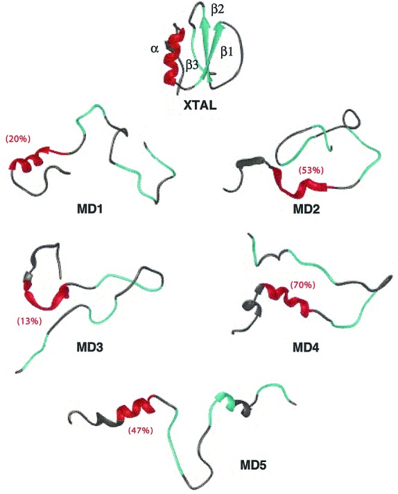
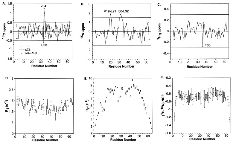
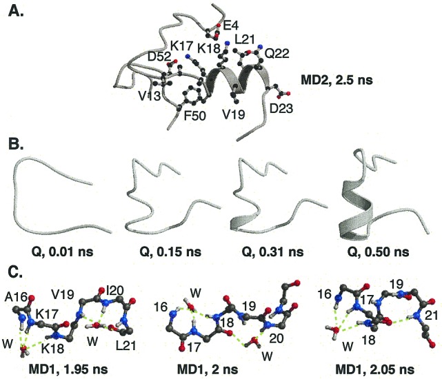
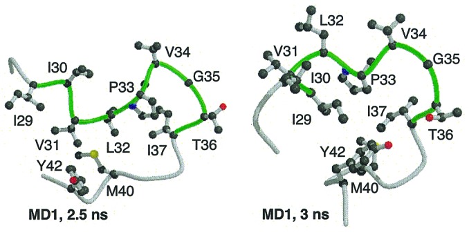
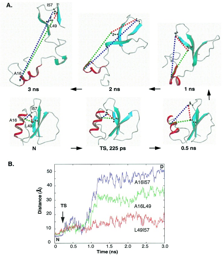
Previous experimental and theoretical studies have produced high-resolution descriptions of the native and folding transition states of chymotrypsin inhibitor 2 (CI2). In similar fashion, here we use a combination of NMR experiments and molecular dynamics simulations to examine the conformations populated by CI2 in the denatured state. The denatured state is highly unfolded, but there is some residual native helical structure along with hydrophobic clustering in the center of the chain. The lack of persistent nonnative structure in the denatured state reduces barriers that must be overcome, leading to fast folding through a nucleation-condensation mechanism. With the characterization of the denatured state, we have now completed our description of the folding/unfolding pathway of CI2 at atomic resolution.


Figures

Figure 1
Figure 1
Crystal structure of CI2 (23) and final structures from each of the denaturation simulations (3 ns, MD1–MD4, 20 ns for MD5). The residues (–23) involved in the native α-helix are colored red. β-Strands 1–3 are colored cyan and are defined as follows: β1, residues 27–34; β2, 46–52; and β3, 59–63. The average helix content of residues 13–23 in each denatured ensemble is given in parentheses (27). Secondary structure, by using the method of Kabsch and Sander (33), is displayed as a ribbon. Fig. 1 was made by using University of California, San Francisco, midasplus (34), and Figs. 3–5 were created with molscript (35) and raster3d (36).
Figure 2
Figure 2
Structural and dynamic results of NMR experiments of denatured CI2. The deviation of the chemical shifts from random coil values for denatured CI2: (A) 13C, (B) 15N, and (C) 1H. Relaxation rates and correlation times for denatured CI2: (D) R1, (E) R2 and (F) {1H-15N} NOE values. Plots of the correlation times (τc) for denatured CI2 parallel those of the R2 values.
Figure 3
Figure 3
Residual helical structure in the denatured state. (A) Residual helix in the 2.5-ns structure from the MD2 simulation. The hydrophobic side chains of the helix are protected from solvent by the rest of the denatured protein. The more hydrophilic side chains of Lys-17 and -18 form salt bridges with acidic side chains allowing their hydrophobic methylene side chains to cluster away from solvent. (B) Residues 11 (Top) to 26 from the Q simulation. Native helix formed between residues 17–22. (C) Mechanism of the formation of the initial helical turn in the denatured state. In MD1, two water molecules form hydrogen bonds with the main chain amide hydrogens of residues 17–21. One water molecule initiated a turn-like structure between residues 16–18, whereas the other led to a turn-like structure between residues 19–21. The two water molecules ordered the region for helical formation. At 2.05 ns, the carbonyl oxygens of residues 16 and 17 formed i → i + 4 hydrogen bonds with residues 20 and 21, expelling the water from the region.
Figure 4
Figure 4
Hydrophobic clustering near the center of the sequence. The main chain of residues 27–43 is presented with residues 29–37 in green.
Figure 5
Figure 5
The folding nucleus comes together only in the transition state. (A) Snapshots from MD1 highlighting the nucleation-site region, Ala-16, Leu-49, and Ile-57. These residues are tightly packed in the native structure and retain some contacts through, but not beyond, the transition state. (B) Distances between the three residues as a function of simulation time.

References

    1. Wong K B, Clarke J, Bond C J, Neira J L, Freund S M V, Fersht A R F, Daggett V. J Mol Biol. 2000;296:1257–1282. - PubMed
    1. Matouschek A, Kellis J T, Jr, Serrano L, Fersht A R. Nature (London) 1989;340:122–126. - PubMed
    1. Li A, Daggett V. Proc Natl Acad Sci USA. 1994;91:10430–10434. - PMC - PubMed
    1. Li A, Daggett V. J Mol Biol. 1996;257:412–429. - PubMed
    1. Daggett V, Li A, Itzhaki L S, Otzen D E, Fersht A R. J Mol Biol. 1996;257:430–440. - PubMed

Publication types

LinkOut - more resources