. 2001 Apr 10;98(8):4540-5.
doi: 10.1073/pnas.071375898. Epub 2001 Mar 27.

Galactosides in the rhizosphere: utilization by Sinorhizobium meliloti and development of a biosensor

Affiliations

Galactosides in the rhizosphere: utilization by Sinorhizobium meliloti and development of a biosensor

R M Bringhurst et al. Proc Natl Acad Sci U S A. .

Abstract

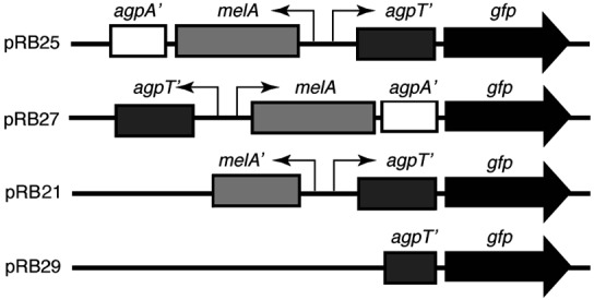
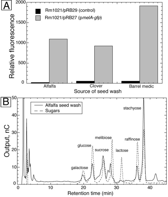
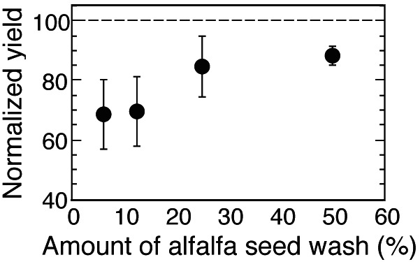
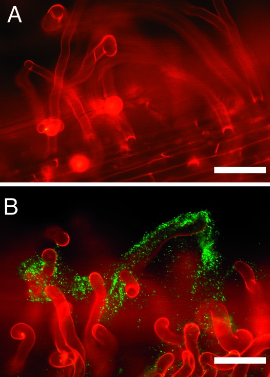
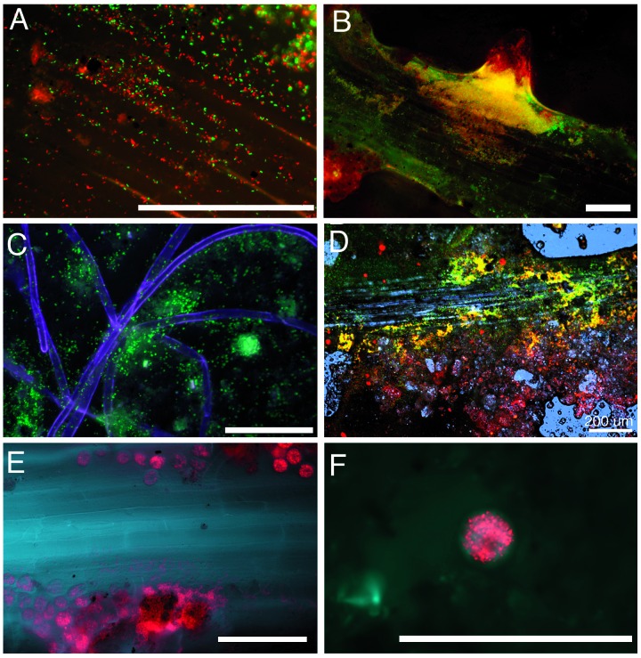
Identifying the types and distributions of organic substrates that support microbial activities around plant roots is essential for a full understanding of plant-microbe interactions and rhizosphere ecology. We have constructed a strain of the soil bacterium Sinorhizobium meliloti containing a gfp gene fused to the melA promoter which is induced on exposure to galactose and galactosides. We used the fusion strain as a biosensor to determine that galactosides are released from the seeds of several different legume species during germination and are also released from roots of alfalfa seedlings growing on artificial medium. Galactoside presence in seed wash and sterile root washes was confirmed by HPLC. Experiments examining microbial growth on alpha-galactosides in seed wash suggested that alpha-galactoside utilization could play an important role in supporting growth of S. meliloti near germinating seeds of alfalfa. When inoculated into microcosms containing legumes or grasses, the biosensor allowed us to visualize the localized presence of galactosides on and around roots in unsterilized soil, as well as the grazing of fluorescent bacteria by protozoa. Galactosides were present in patches around zones of lateral root initiation and around roots hairs, but not around root tips. Such biosensors can reveal intriguing aspects of the environment and the physiology of the free-living soil S. meliloti before and during the establishment of nodulation, and they provide a nondestructive, spatially explicit method for examining rhizosphere soil chemical composition.


Figures

Figure 1
Figure 1
Maps of the gfp fusion plasmids used in this study.
Figure 2
Figure 2
(A) Relative fluorescence of strain Rm1021/pRB29 and strain Rm1021/pRB27 grown in M9 medium with seed wash from three legumes as carbon sources. (B) HPLC chromatogram of alfalfa seed wash overlaid on a chromatogram of a mixture of seven sugars.
Figure 3
Figure 3
Mean (± SD) growth yield of strain RB6 (agpT-) grown using various concentrations of alfalfa seed wash as a carbon source (n = 4). Yields of strain RB6 (agpT-) are normalized to the yields obtained by the wild-type strain Rm1021. The dashed line shows the value at which the yield of strain RB6 would be equal to the yield of strain Rm1021.
Figure 4
Figure 4
Induction of the pmelA-gfp reporter in the rhizosphere of alfalfa seedlings grown in artificial medium. (A) Fluorescent micrograph of the control strain Rm1021/pRB29 inoculated onto the roots of alfalfa seedlings grown on BNM. (B) Fluorescent micrograph of the pmelA-gfp reporter strain Rm1021/pRB27 inoculated onto the roots of alfalfa seedlings grown on BNM. (Bars = 100 μm.)
Figure 5
Figure 5
Induction of the pmelA-gfp reporter in the rhizosphere of legumes and grasses growing in nonsterilized soil. All panels except C show roots in microcosms inoculated with a mixture of Rm1021/pRB27 (pmelAgfp) and Rm1021/pDG77 (constitutive DsRed). C shows root hairs in a microcosm inoculated with Rm1021/pRB27 (pmelAgfp) only. In all micrographs, green fluorescence (indicating the presence of galactosides) is colored green, red fluorescence from DsRed is colored red, and in C, E, and F, root autofluorescence (visualized with a brief pulse of UV light) is colored blue. Root autofluorescence was not imaged separately when roots in A and B were examined. Colocalized green and red fluorescence combine to give yellow. [Bars = 100 μm (except in D).] (A) Red and green fluorescent bacteria on the root surface of an Avena sativa seedling. (B) Alfalfa root with red and green fluorescent bacteria. Colocalized red and green bacteria are especially prevalent around an emerging lateral root. (C) Green fluorescent bacteria associated with root hairs of Bromus hordeaceus. (D) Confocal micrograph of an alfalfa root in soil; blue represents reflected confocal laser light revealing root cells, soil structure, and water droplets. (E) Zone of an alfalfa root where protozoa are grazing on red bacteria. (F) Close-up of another alfalfa root showing individual round protozoa with numerous red fluorescent bacteria inside.

References

    1. Mylona P, Pawlowski K, Bisseling T. Plant Cell. 1995;7:869–885. - PMC - PubMed
    1. Spaink H P. Annu Rev Phytopathol. 1995;33:345–368. - PubMed
    1. Hirsch A M. Curr Opin Plant Biol. 1999;2:320–326. - PubMed
    1. Gray K M, Pearson J P, Downie J A, Boboye B E A, Greenberg E P. J Bacteriol. 1996;178:372–376. - PMC - PubMed
    1. Triplett E, Sadowsky M J. Annu Rev Microbiol. 1992;46:399–428. - PubMed

Publication types

LinkOut - more resources