. 2001 Apr 10;98(8):4776-81.
doi: 10.1073/pnas.081072398. Epub 2001 Mar 27.

Ca2+-binding activity of a COOH-terminal fragment of the Drosophila BK channel involved in Ca2+-dependent activation

Affiliations

Ca2+-binding activity of a COOH-terminal fragment of the Drosophila BK channel involved in Ca2+-dependent activation

S Bian et al. Proc Natl Acad Sci U S A. .

Abstract

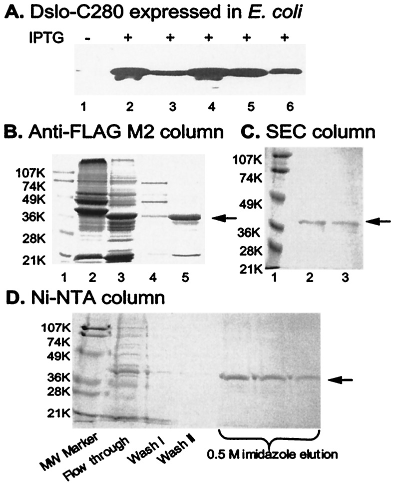
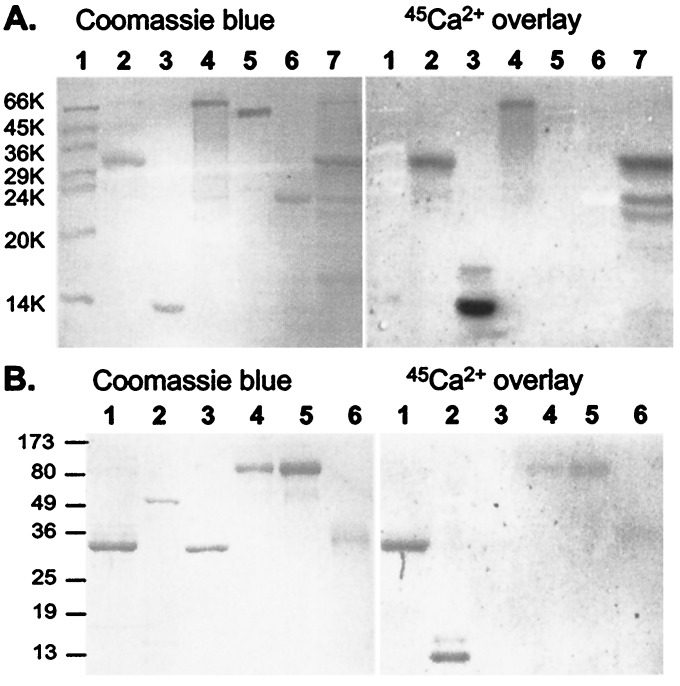
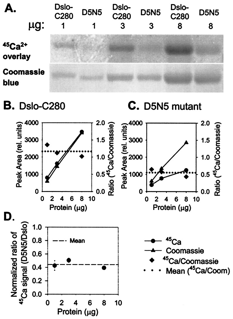
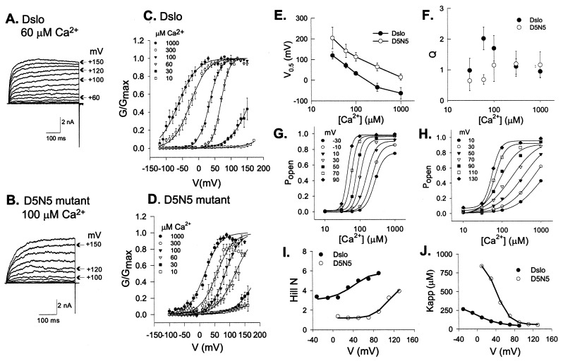
Mutational and biophysical analysis suggests that an intracellular COOH-terminal domain of the large conductance Ca(2+)-activated K(+) channel (BK channel) contains Ca(2+)-binding site(s) that are allosterically coupled to channel opening. However the structural basis of Ca(2+) binding to BK channels is unknown. To pursue this question, we overexpressed the COOH-terminal 280 residues of the Drosophila slowpoke BK channel (Dslo-C280) as a FLAG- and His(6)-tagged protein in Escherichia coli. We purified Dslo-C280 in soluble form and used a (45)Ca(2+)-overlay protein blot assay to detect Ca(2+) binding. Dslo-C280 exhibits specific binding of (45)Ca(2+) in comparison with various control proteins and known EF-hand Ca(2+)-binding proteins. A mutation (D5N5) of Dslo-C280, in which five consecutive Asp residues of the "Ca-bowl" motif are changed to Asn, reduces (45)Ca(2+)-binding activity by 56%. By electrophysiological assay, the corresponding D5N5 mutant of the Drosophila BK channel expressed in HEK293 cells exhibits lower Ca(2+) sensitivity for activation and a shift of approximately +80 mV in the midpoint voltage for activation. This effect is associated with a decrease in the Hill coefficient (N) for activation by Ca(2+) and a reduction in apparent Ca(2+) affinity, suggesting the loss of one Ca(2+)-binding site per monomer. These results demonstrate a functional correlation between Ca(2+) binding to a specific region of the BK protein and Ca(2+)-dependent activation, thus providing a biochemical approach to study this process.


Figures

Figure 1
Figure 1
Schematic overview of Drosophila BK channel and expressed fragment. (A) Topology diagram of Dslo α subunit showing the location of Core and Tail domains. (B) Construction of Dslo-C280 protein (and D5N5 mutant) expressed in E. coli. (C) Sequence of Ca-bowl motif (Dslo residues T952-Q979) of Dslo-C280 and D5N5 mutant.
Figure 2
Figure 2
Expression and purification of Dslo-C280. (A) Immunoblot detection of Dslo-C280 band by anti-FLAG M2 antibody in various fractions of E. coli before (lane 1) or after (lanes 2–6) induction with IPTG. Lanes: 1, whole cells before IPTG; 2, insoluble protein pellet from lysed cells; 3, soluble protein from lysed cells; 4, whole cells; 5, soluble fraction after high sucrose treatment; and 6, soluble fraction after osmotic shock. (B) Purification via FLAG epitope on M2 anti-FLAG affinity column. SDS/PAGE of various fractions. Lanes: 1, molecular mass markers (K = kDa); 2, crude soluble extract; 3, crude inclusion body sample; 4, fraction from soluble extract after M2 column purification; and 5, fraction of renatured inclusion protein after M2 column purification. (C) Purification by SEC. SDS/PAGE of fractions from TSK G3000SW column. Lanes: 1, molecular mass markers; 2, SEC peak fraction at ≈68 kDa; and 3, SEC peak fraction at ≈34 kDa. (D) Purification via His6-tag of renatured inclusion protein. SDS/PAGE of various fractions from Ni2+-NTA column. The arrow in B, C, and D points to the Dslo-C280 band.
Figure 3
Figure 3
Protein blot assays demonstrating specificity of 45Ca2+ binding. Purified Dslo-C280 and various control proteins were subjected to SDS/PAGE, electroblotted onto poly(vinylidene difluoride) membrane, and processed for 45Ca2+-overlay assay: (Right) image of exposed film; (Left) same blot stained with Coomassie Blue. (A) Lanes: 1, molecular mass markers; 2, Dslo-C280 (≈7 μg); 3, bovine calmodulin (4 μg); 4, rabbit calpain (6 μg); 5, FLAG-tagged E. coli alkaline phosphatase (4 μg); 6; bovine trypsin (4 μg); and 7, crude extract from E. coli expressing Dslo-C280. (B) Lanes: 1, Dslo-C280 (3 μg); 2, chicken troponin complex (6 μg); 3, human annexin V (3 μg); 4, Aspergillus niger glucose oxidase (3 μg); 5, glucose oxidase (6 μg); and 6, chicken ovomucoid protease inhibitor (3 μg).
Figure 4
Figure 4
Comparison of 45Ca2+-binding activity of Dslo-C280 and corresponding D5N5 mutation. (A) Purified Dslo-C280 and D5N5 mutant (1, 3, and 8 μg) were subjected to SDS/PAGE, electroblotted onto PDVF, and assayed by 45Ca2+-overlay method: (Upper) image of exposed film; (Lower) Coomassie blue-stained protein bands. (B and C) Densitometric measurement of protein bands (▴), 45Ca2+ signal (●), and 45Ca2+/Coomassie ratio (♦) for Dslo-C280 (B) and D5N5 mutant (C). (D) Protein-normalized ratio of 45Ca2+-binding activity of D5N5 mutant relative to Dslo-C280.
Figure 5
Figure 5
Analysis of the effect of the D5N5 Ca-bowl mutation on BK channel activation. Dslo and D5N5 mutant BK channels were expressed in HEK293 cells and characterized by patch clamp analysis in inside-out patches. (A and B) Current traces evoked by consecutive voltage steps of +10 mV from a holding potential of −140 mV for Dslo (A) and −120 mV for D5N5 (B) with 60 μM and 100 μM Ca2+ in the bath solution, respectively. (C and D) Normalized G-V curves representing the average of 5 to 12 patches at indicated Ca2+ concentrations for parent Dslo (C) and D5N5 mutant (D). Error bars denote ±SEM. Solid curves are fits to a Boltzmann relationship (Eq. 1). (E and F) Comparison of V0.5 (E) and Q (F) vs. [Ca2+]. Data points are the mean ±SD of Boltzmann fit parameters from 5–12 patches. (G and H) Mean values of steady-state Popen (G/Gmax) as a function of [Ca2+] at various voltages compared for Dslo (G) and D5N5 mutant (H). Solid curves indicate fits to Hill equation (Eq. 2). (I and J) Comparison of Hill parameters, N (I) and Kapp (J) vs. voltage.

References

    1. Toro L, Wallner M, Meera P, Tanaka Y. News Physiol Sci. 1998;13:112–117. - PubMed
    1. Meera P, Wallner M, Jiang Z, Toro L. FEBS Lett. 1996;382:84–88. - PubMed
    1. Cui J, Cox D H, Aldrich R W. J Gen Physiol. 1997;109:647–673. - PMC - PubMed
    1. Butler A, Tsunoda S, McCobb D P, Wei A, Salkoff L. Science. 1993;261:221–224. - PubMed
    1. Meera P, Wallner M, Song M, Toro L. Proc Natl Acad Sci USA. 1997;94:14066–14071. - PMC - PubMed

Publication types

Substances

LinkOut - more resources