. 2001 Apr 10;98(8):4492-7.
doi: 10.1073/pnas.081076898. Epub 2001 Mar 27.

Mammalian mad2 and bub1/bubR1 recognize distinct spindle-attachment and kinetochore-tension checkpoints

Affiliations

Mammalian mad2 and bub1/bubR1 recognize distinct spindle-attachment and kinetochore-tension checkpoints

D A Skoufias et al. Proc Natl Acad Sci U S A. .

Abstract

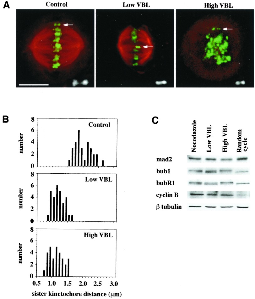
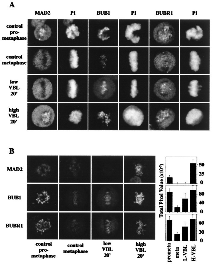
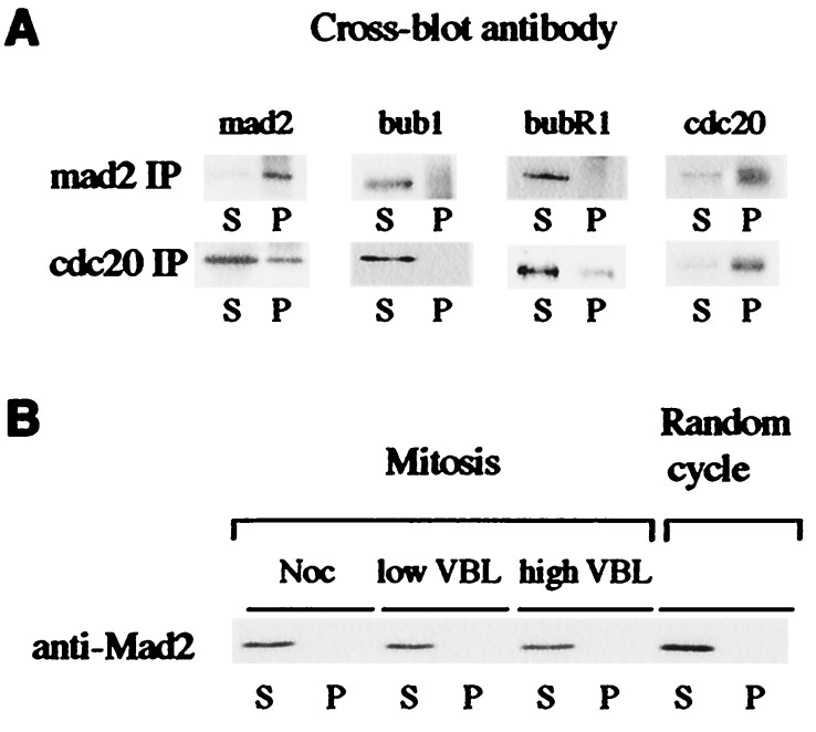
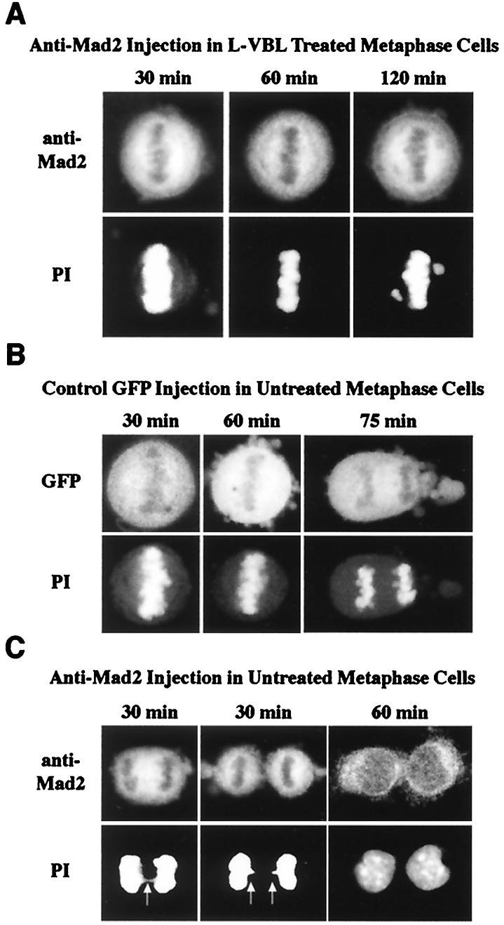
Metaphase checkpoint controls sense abnormalities of chromosome alignment during mitosis and prevent progression to anaphase until proper alignment has been attained. A number of proteins, including mad2, bub1, and bubR1, have been implicated in the metaphase checkpoint control in mammalian cells. Metaphase checkpoints have been shown, in various systems, to read loss of either spindle tension or microtubule attachment at the kinetochore. Characteristically, HeLa cells arrest in metaphase in response to low levels of microtubule inhibitors that leave an intact spindle and a metaphase plate. Here we show that the arrest induced by nanomolar vinblastine correlates with loss of tension at the kinetochore, and that in response the checkpoint proteins bub1 and bubR1 are recruited to the kinetochore but mad2 is not. mad2 remains competent to respond and is recruited at higher drug doses that disrupt spindle association with the kinetochores. Further, although mad2 forms a complex with cdc20, it does not associate with bub1 or bubR1. We conclude that mammalian bub1/bubR1 and mad2 operate as elements of distinct pathways sensing tension and attachment, respectively.


Figures

Figure 1
Figure 1
Treatment of HeLa cells with low concentrations of vinblastine suppresses microtubule dynamics while maintaining a bipolar mitotic spindle and attached kinetochores. (A) Double-label immunofluorescence microscopy for centromeres (green; detected with CREST variant scleroderma human autoimmune serum) and antitubulin (red) demonstrates that 6.7 nM VBL does not perturb bipolar spindle association with kinetochores, as compared with untreated control metaphase cells. By contrast, 0.5 μM VBL induces complete disassembly of the mitotic spindle. (Bar = 10 μm.) Insets show enlargements of kinetochore pairs representative of those used for measurements. Point-to-point measurements were made by using comos software. Kinetochore pairs were selected by their close proximity, presence in the same focal plane, and, when at metaphase, alignment with the long axis of the spindle. Measured distances of the Inset kinetochores shown were: control, 2.04 μm; L-VBL, 1.24 μm; H-VBL, 1.16 μm. (B) VBL (6.7 nM) suppresses spindle tension at kinetochores, reducing the distance between sister kinetochores relative to that of untreated control metaphases (from 1.95 ± 0.28 μm to 1.15 ± 0.2 μm), yielding values comparable to those in 0.5 μM VBL-treated cells with no spindles (1.1 ± 0.23 μm). (C) The whole cell levels of mad2, bub1, and bubR1, as determined by immunoblotting, remain constant in mitotic cells in which spindle dynamics are suppressed (6.7 nM VBL) or in which the spindle is disassembled (0.5 μM VBL or 1.7 μM nocodazole). Cyclin B blots are shown to confirm mitotic status. β-tubulin blots are shown to confirm equal loading of all extracts.
Figure 2
Figure 2
bub1 and bubR1, but not mad2, respond to the absence of spindle tension by binding to kinetochores. (A) mad2 is absent from kinetochores that have attached microtubules and is insensitive to loss of tension by the addition of 6.7 nM VBL for 20 min. In contrast, mad2 reassociates with kinetochores when attachment is abolished by the addition of 0.5 μM VBL. bub1 and bubR1 are minimally detectable at kinetochores of control metaphase cells but reassociate at kinetochores of metaphase arrays when tension is suppressed by the addition of 6.7 nM VBL or if microtubule attachment is abolished by the addition of 0.5 μM VBL. Results were consistent in several independent experiments and (for bub1) with use of two distinct antibodies (data not shown). For each antigen, all images were collected with a confocal microscope by using constant settings established by imaging control metaphases. (Bar = 5 μm.) (B) Levels of antigens were quantitated for different conditions by using the photon counting mode of the confocal microscope. (Left) The images shown were collected in photon-counting mode at identical machine settings for each antigen. (Right) Compiled data for each of the different conditions with at least 300 kinetochores counted for each data set. Standard deviations were as indicated.
Figure 3
Figure 3
Associations between mad2, bub1, bubR1, and cdc20. (A) Western blots showing neither bub1 nor bubR1 coimmunoprecipitates with mad2 in extracts from mitotic cells (derived by shakeoff after 4-h blockage with nocodazole). Cdc20 coimmunoprecipitates with mad2 and weakly with bubR1 but not with bub1. (B) Western blot showing mad2 is entirely soluble in cell extracts. Supernatants (S) and pellets (P) were loaded on the basis of equivalent initial volumes.
Figure 4
Figure 4
Response of VLB-treated HeLa to anti-mad2 microinjection. Immunofluorescence micrographs show the result of microinjection of either GFP or anti-mad2 antibody. (A) Cells pretreated for 20 min with low VBL (L-VBL) remain in metaphase configuration for at least 2 h (the last time point taken) after microinjection of anti-mad2 antibody. (B) GFP microinjection was performed as a positive control in untreated metaphase cells. Shown are representative cells that remain at metaphase for 30 and 60 min, respectively, after microinjection. At later times (an anaphase at 75 min is shown here), cells progress to anaphase and exit mitosis. (C) Anti-mad2 microinjection of untreated metaphase cells. In contrast to GFP controls, there is a rapid progression through anaphase and cleavage. Representative cells are shown here 30 min after microinjection in late anaphase and telophase, respectively. The anaphase cells both show evidence of chromosome bridging (arrows), indicating premature entry into anaphase (the propidium iodide stain in both 30-min images has been enhanced to make the bridging visible). GFP or anti-mad2 labels positively identify those cells that had been successfully microinjected. PI, propidium iodide stain to assess the status of the chromosomes after microinjection.

References

    1. Li R, Murray A W. Cell. 1991;66:519–531. - PubMed
    1. Hoyt M A, Totis L, Roberts B T. Cell. 1991;66:507–517. - PubMed
    1. Amon A. Curr Opin Genet Dev. 1999;9:69–75. - PubMed
    1. Li Y, Benezra R. Science. 1996;274:246–248. - PubMed
    1. Chen R H, Waters J C, Salmon E D, Murray A W. Science. 1996;274:242–246. - PubMed

Publication types

LinkOut - more resources