. 2001 Mar 27;98(7):3796-801.
doi: 10.1073/pnas.071245198.

Local osmotic gradients drive the water flux associated with Na(+)/glucose cotransport

Affiliations

Local osmotic gradients drive the water flux associated with Na(+)/glucose cotransport

P P Duquette et al. Proc Natl Acad Sci U S A. .

Abstract

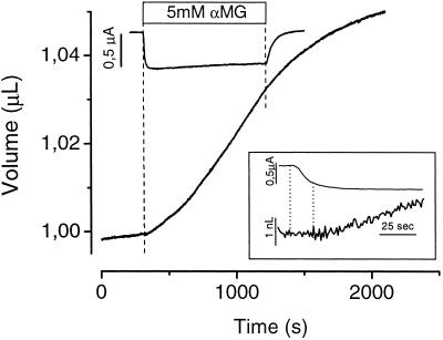
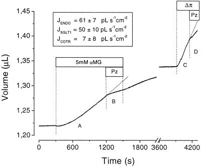
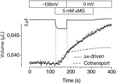
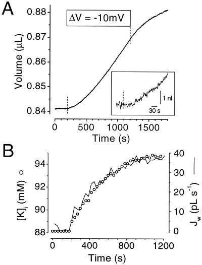
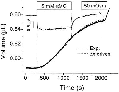
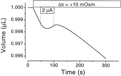
It recently was proposed [Loo, D. D. F., Zeuthen, T., Chandy, G. & Wright, E. M. (1996) Proc. Natl. Acad. Sci. USA 93, 13367--13370] that SGLT1, the high affinity intestinal and renal sodium/glucose cotransporter carries water molecules along with the cosubstrates with a strict stoichiometry of two Na(+), one glucose, and approximately 220 water molecules per transport cycle. Using electrophysiology together with sensitive volumetric measurements, we investigated the nature of the driving force behind the cotransporter-mediated water flux. The osmotic water permeability of oocytes expressing human SGLT1 (L(p) +/- SE) averaged 3.8 +/- 0.3 x 10(-4) cm x s(-1) (n = 15) and addition of 100 microM phlorizin (a specific SGLT1 inhibitor) reduced the permeability to 2.2 +/- 0.2 x 10(-4) cm x s(-1) (n = 15), confirming the presence of a significant water permeability closely associated with the cotransporter. Addition of 5 mM alpha-methyl-glucose (alpha MG) induced an average inward current of 800 +/- 10 nA at -50 mV and a water influx reaching 120 +/- 20 pL cm(-2) x s(-1) within 5-8 min. After rapidly inhibiting the Na(+)/glucose cotransport with phlorizin, the water flux remained significantly elevated, clearly indicating the presence of a local osmotic gradient (Delta pi) estimated at 16 +/- 2 mOsm. In short-term experiments, a rapid depolarization from -100 to 0 mV in the presence of alpha MG decreased the cotransport current by 94% but failed to produce a comparable reduction in the swelling rate. A mathematical model depicting the intracellular accumulation of transported osmolytes can accurately account for these observations. It is concluded that, in SGLT1-expressing oocytes, alpha MG-dependent water influx is induced by a local osmotic gradient by using both endogenous and SGLT1-dependent water permeability.


Figures

Figure 1
Figure 1
αMG-dependent water flux. Oocyte current and volume are displayed as a function of time. From t = 300–1,200 s, 5 mM αMG was added to the bathing solution. (Inset) Enlargement of the initial portion of the curves. Dotted lines, represent the time needed to reach 80% of the final cotransport current.
Figure 2
Figure 2
Analysis of water transport during prolonged exposure to 5 mM αMG. Oocyte volume is displayed as a function of time. αMG was present between t = 300 s and 1,500 s, and Pz was added as indicated. After a recovery period in the absence of αMG, an osmotic shock was imposed to enable water permeability measurement. (Inset) Calculated fluxes passing through the three presumed water pathways: endogenous water permeability (JENDO), SGLT1 passive permeability (JSGLT1), and cotransported with Na+ and αMG (JCOTR).
Figure 3
Figure 3
Effect of a rapid reduction in the cotransported current. After a stabilization period at a membrane potential of −100 mV, 5 mM αMG is introduced in the bathing solution. After establishment of a steady-state cotransport current (a 60-s period), the membrane potential was stepped to 0 mV to abruptly reduce the cotransport current. At t = 300 s, αMG was removed to allow a determination of background current at 0 mV. The volume data were either fitted according to the water-cotransport hypothesis (240 water molecules/αMG molecule, dashed line) or by using the osmolyte accumulation model (full line).
Figure 4
Figure 4
Effect of a K+ inward current on oocyte volume and intracellular K+ concentration. (A) Average volume measurements in ROMK2-injected oocytes (n = 3). At t = 180 s, the oocyte membrane was hyperpolarized by 10 mV to stimulate an inward K current averaging 1.8 μA. (Inset) Enlargement of the initial portion of the curve shown in A. The dotted line represents the time at which the inward current was stimulated. (B) Parallel increases in intracellular K+ concentration ([K]i, ○) and in the instantaneous water flux (Jw, full line). [K]i was obtained from the K+ reversal potential and Jw was obtained from the slope of the curve shown in A, which was measured every 30 s using a time window of ± 25 s.
Figure 5
Figure 5
Theoretical fit using the osmolyte accumulation model. Oocyte current and volume are shown as a function of time. From t = 300–1,200 s, 5 mM αMG was added to the bathing solution and at t = 2,100 s, 50 mM mannitol was removed from the external solution. Superposition of the experimental data (full line) to the theoretical prediction (dashed line) from the accumulation model are presented.
Figure 6
Figure 6
Passive water transport against an apparent osmotic gradient. The osmolyte accumulation model was used to calculate the oocyte volume as a function of time after presenting an hypertonic (+10 mOsm) bath solution. The parameters used were: Lp = 7 × 10⁻⁴ cm⋅s−1, D = 0.5 × 10⁻⁶ cm2⋅s−1, and n = 5 osmolytes/αMG transported. From t = 50–100 s, a 2 μA inward cotransport current is applied.

Comment in

References

    1. Finkelstein A. Water Movements Through Lipid Bilayers, Pores, and Plasma Membranes: Theory and Reality. New York: Wiley; 1987.
    1. Fischbarg J, Kuang K Y, Vera J C, Arant S, Silverstein S C, Loike J, Rosen O M. Proc Natl Acad Sci USA. 1990;87:3244–3247. - PMC - PubMed
    1. Agre P, Preston G M, Smith B L, Jung J S, Raina S, Moon C, Guggino W B, Nielsen S. Am J Physiol. 1993;265:F463–F476. - PubMed
    1. Loike J D, Hickman S, Kuang K, Xu M, Cao L, Vera J C, Silverstein S C, Fischbarg J. Am J Physiol. 1996;271:C1774–C1779. - PubMed
    1. Loo D D F, Zeuthen T, Chandy G, Wright E M. Proc Natl Acad Sci USA. 1996;93:13367–13370. - PMC - PubMed

Publication types

MeSH terms