. 2001 Mar 27;98(7):3826-31.
doi: 10.1073/pnas.071043198.

Switch from Myc/Max to Mad1/Max binding and decrease in histone acetylation at the telomerase reverse transcriptase promoter during differentiation of HL60 cells

Affiliations

Switch from Myc/Max to Mad1/Max binding and decrease in histone acetylation at the telomerase reverse transcriptase promoter during differentiation of HL60 cells

D Xu et al. Proc Natl Acad Sci U S A. .

Abstract

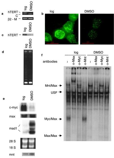
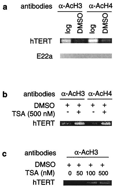
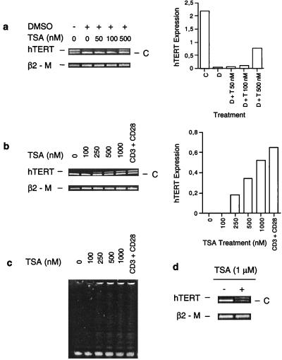
Recent evidence suggests that the Myc and Mad1 proteins are implicated in the regulation of the gene encoding the human telomerase reverse transcriptase (hTERT), the catalytic subunit of telomerase. We have analyzed the in vivo interaction between endogenous c-Myc and Mad1 proteins and the hTERT promoter in HL60 cells with the use of the chromatin immunoprecipitation assay. The E-boxes at the hTERT proximal promoter were occupied in vivo by c-Myc in exponentially proliferating HL60 cells but not in cells induced to differentiate by DMSO. In contrast, Mad1 protein was induced and bound to the hTERT promoter in differentiated HL60 cells. Concomitantly, the acetylation of the histones at the promoter was significantly reduced. These data suggest that the reciprocal E-box occupancy by c-Myc and Mad1 is responsible for activation and repression of the hTERT gene in proliferating and differentiated HL60 cells, respectively. Furthermore, the histone deacetylase inhibitor trichostatin A inhibited deacetylation of histones at the hTERT promoter and attenuated the repression of hTERT transcription during HL60 cell differentiation. In addition, trichostatin A treatment activated hTERT transcription in resting human lymphocytes and fibroblasts. Taken together, these results indicate that acetylation/deacetylation of histones is operative in the regulation of hTERT expression.


Figures

Figure 1
Figure 1
Down-regulation of hTERT/telomerase activity is closely associated with changes in c-Myc and Mad1 expression during the differentiation of HL60 cells. HL60 cells were treated with DMSO for 48 h and analyzed for (a) hTERT mRNA expression by competitive RT-PCR; (b and c) expression of hTERT protein by immunofluorescence and Western blot; (d) telomerase activity by telomeric repeat amplification protocol assay; (e) c-Myc, Max, and Mad1 expression by Northern blot (Top), the ethidium bromide-stained gel visualizing the 18 and 28 S RNA (Middle), and Mnt expression by RT-PCR (Bottom); and (f) EMSA demonstrating DNA binding activities to the hTERT E-box in HL60 extracts. Antibodies used for supershifts are indicated at the top, and protein complexes binding to the hTERT oligo are indicated to the left. Log, logarithmically growing cells; DMSO, DMSO-treated (differentiated) cells; C, competitor for hTERT PCR product; β2-M, β2-microglobulin, internal control for the hTERT and Mnt RT-PCR.
Figure 2
Figure 2
In vivo binding of c-Myc/Max, Mad1/Max, Mnt/Max, and USF to the hTERT proximal promoter in HL60 cells. A ChIP assay was performed on logarithmically growing and DMSO-treated HL60 cells, and the precipitated chromatin was PCR-amplified with the use of specific primers. (a) (Left) Schematic presentation of E-boxes (□) in the hTERT promoter and in the E22a locus, and the location of the respective primer sequences (■) for PCR analysis. The numbers below the hTERT promoter indicate the position of the PCR primers relative to ATG. The size of the E22a PCR product is shown. (Right) In vivo identification of reciprocal E-box occupancy by c-Myc, Max, and Mad1 at the hTERT promoter in logarithmically growing (log) and differentiated (DMSO) HL60 cells. β-Galactosidase antibodies were used as controls. Input: PCRs performed on total chromatin from differentiated HL60 cells. There is an absence of c-Myc, Max, and Mad1 binding to the control E22a locus that contains E-boxes but is not transcribed. (b) Absence of USF and presence of Mnt at the hTERT promoter in vivo in undifferentiated (−) and DMSO-differentiated (+) HL60 cells. Neither Mnt nor USF was present at the control E22a locus.
Figure 3
Figure 3
Changes in histone acetylation at the hTERT promoter and TSA-mediated hyperacetylation of histones during HL60 differentiation. A ChIP assay was performed on logarithmically growing and DMSO-treated HL60 cells as described in Materials and Methods. (a) A decrease in acetylation of H3 and H4 histones at the hTERT promoter in differentiated HL60 cells. The absence of acetylated histones at the E22a fragment that contains an E-box but is not transcribed. Log, logarithmically growing cells; DMSO, DMSO-treated (differentiated) cells. (b) Abolishment of the differentiation-associated deacetylation of histones H3 and H4 at the hTERT promoter by TSA treatment in HL60 cells. Cells were induced to differentiate by DMSO overnight in the absence or presence of TSA as indicated. (c) TSA-mediated dose-dependent accumulation of histone H3 at the hTERT promoter in differentiating HL60 cells. Cells were treated with DMSO overnight in the absence or presence of various concentrations of TSA as indicated.
Figure 4
Figure 4
Inhibition of HDACs by TSA attenuates down-regulation of hTERT mRNA expression in differentiated HL60 cells and activates hTERT transcription and telomerase in resting human T cells and fibroblasts. C, Competitor for hTERT fragment; β2-M, β2-microglobulin, internal control for the RT-PCR. (a) (Left) Competitive RT-PCR for hTERT mRNA in HL60 cells treated with TSA and/or DMSO. HL60 cells were incubated with TSA at various concentrations for 30 min, followed by overnight culture together with DMSO and analysis for hTERT mRNA expression. (Right) Quantitative expression of hTERT mRNA levels based on the signals to the left. D, DMSO; T, TSA. (b) (Left) Competitive RT-PCR for hTERT mRNA in T cells treated with different concentrations of TSA or anti-CD3 and anti-CD28 antibodies. (Right) Quantitative expression of hTERT mRNA levels based on the signals to the left. (c) Telomerase activity in T cells treated with different concentrations of TSA or anti-CD3 and anti-CD28 antibodies. (d) Competitive RT-PCR for hTERT mRNA in normal human fibroblasts treated with TSA.

References

    1. Henriksson M, Lüscher B. Adv Cancer Res. 1996;68:109–182. - PubMed
    1. Gregor P D, Sawadogo M, Roeder R G. Genes Dev. 1990;4:1730–1740. - PubMed
    1. McMahon S, Wood M A, Cole M D. Mol Cell Biol. 2000;20:556–562. - PMC - PubMed
    1. Cole M D, McMahon S B. Oncogene. 1999;18:2916–2924. - PubMed
    1. Bouchard C, Thieke K, Maier A, Saffrich R, Hanley-Hyde J, Ansorge W, Reed S, Sicinski P, Bartek J, Eilers M. EMBO J. 1999;18:5321–5333. - PMC - PubMed

Publication types

MeSH terms

LinkOut - more resources